
|
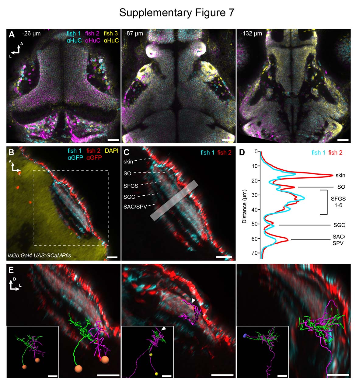
Fig. S7
Accuracy of registration and addition of anatomical reference patterns. (A) Three 5 dpf Optobow-nPA-expressing larvae stained for HuC have been registered into one reference brain. Note the accuracy of registration at different z levels (labeled as distance from dorsal skin in μm). Scale bar, 30 μm. (B) Two 5 dpf larvae expressing GCaMP6s under control of isl2b:Gal4 (stained for GFP) were co-registered into the reference brain. Scale bar, 20 μm. (C) A closeup of the tectal neuropil shows the different innervation strata of RGC axons. SO, stratum opticum; SFGS, stratum fibrosum et griseum superficiale; SGC, stratum griseum centrale; SAC/SPV, stratum album centrale/stratum periventriculare. Scale bar, 20 μm. (D) Intensity profiles through the boxed region in (C) show the alignment accuracy of RGC innervation strata. (E) Examples of three co-registered cell pairs and their dendritic and axonal arborizations in specific tectal layers. Insets taken from Figure 8. Scale bar, 20 μm.