
IMAGE
Fig. 10
- ID
- ZDB-IMAGE-170822-18
- Genes
- Publication
- Kroeger et al., 2017 - The zebrafish kidney mutant zeppelin reveals that brca2/fancd1 is essential for pronephros development
- All Figures
- Figures for Kroeger et al., 2017
Image
|
Figure Caption
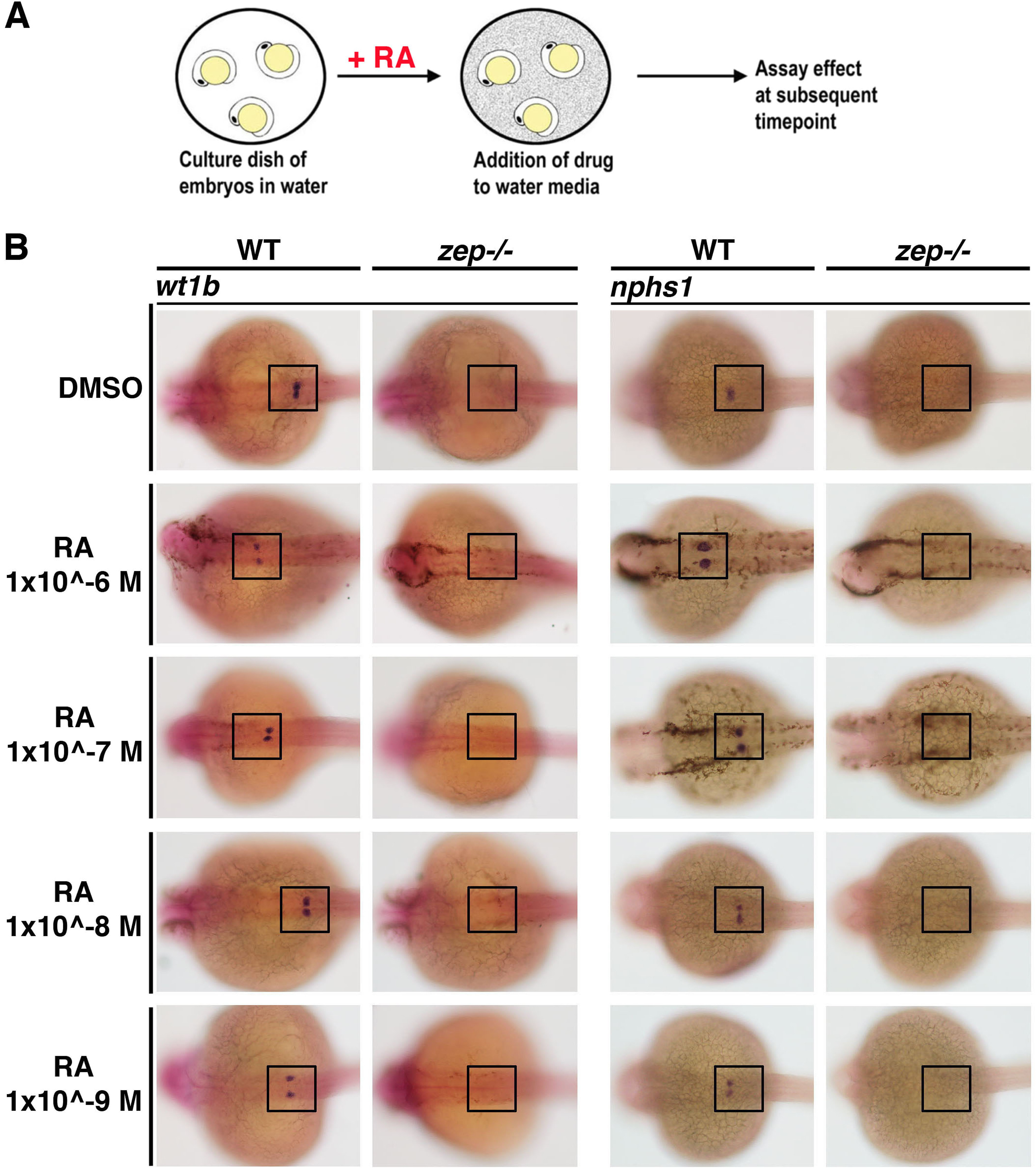
Fig. 10
Addition of exogenous RA during nephrogenesis fails to rescue establishment of the podocyte lineage inzepmutant embryos. (A) Schematic of exogenous RA treatment methodology in zebrafish embryos. (B) While WT siblings exhibit normal podocyte development after RA treatment, zep mutants fail to develop podocytes as assayed by expression of wt1b or nphs1. Embryos are shown in dorsal views, where black boxes demarcate the cervical region where the podocytes develop.
Figure Data
Acknowledgments
This image is the copyrighted work of the attributed author or publisher, and
ZFIN has permission only to display this image to its users.
Additional permissions should be obtained from the applicable author or publisher of the image.
Reprinted from Developmental Biology, 428(1), Kroeger, P.T., Drummond, B.E., Miceli, R., McKernan, M., Gerlach, G.F., Marra, A.N., Fox, A., McCampbell, K.K., Leshchiner, I., Rodriguez-Mari, A., BreMiller, R., Thummel, R., Davidson, A.J., Postlethwait, J., Goessling, W., Wingert, R.A., The zebrafish kidney mutant zeppelin reveals that brca2/fancd1 is essential for pronephros development, 148-163, Copyright (2017) with permission from Elsevier. Full text @ Dev. Biol.