
Fig. 6
- ID
- ZDB-IMAGE-170822-14
- Genes
- Antibodies
- Publication
- Kroeger et al., 2017 - The zebrafish kidney mutant zeppelin reveals that brca2/fancd1 is essential for pronephros development
- All Figures
- Figures for Kroeger et al., 2017
|
Fig. 6
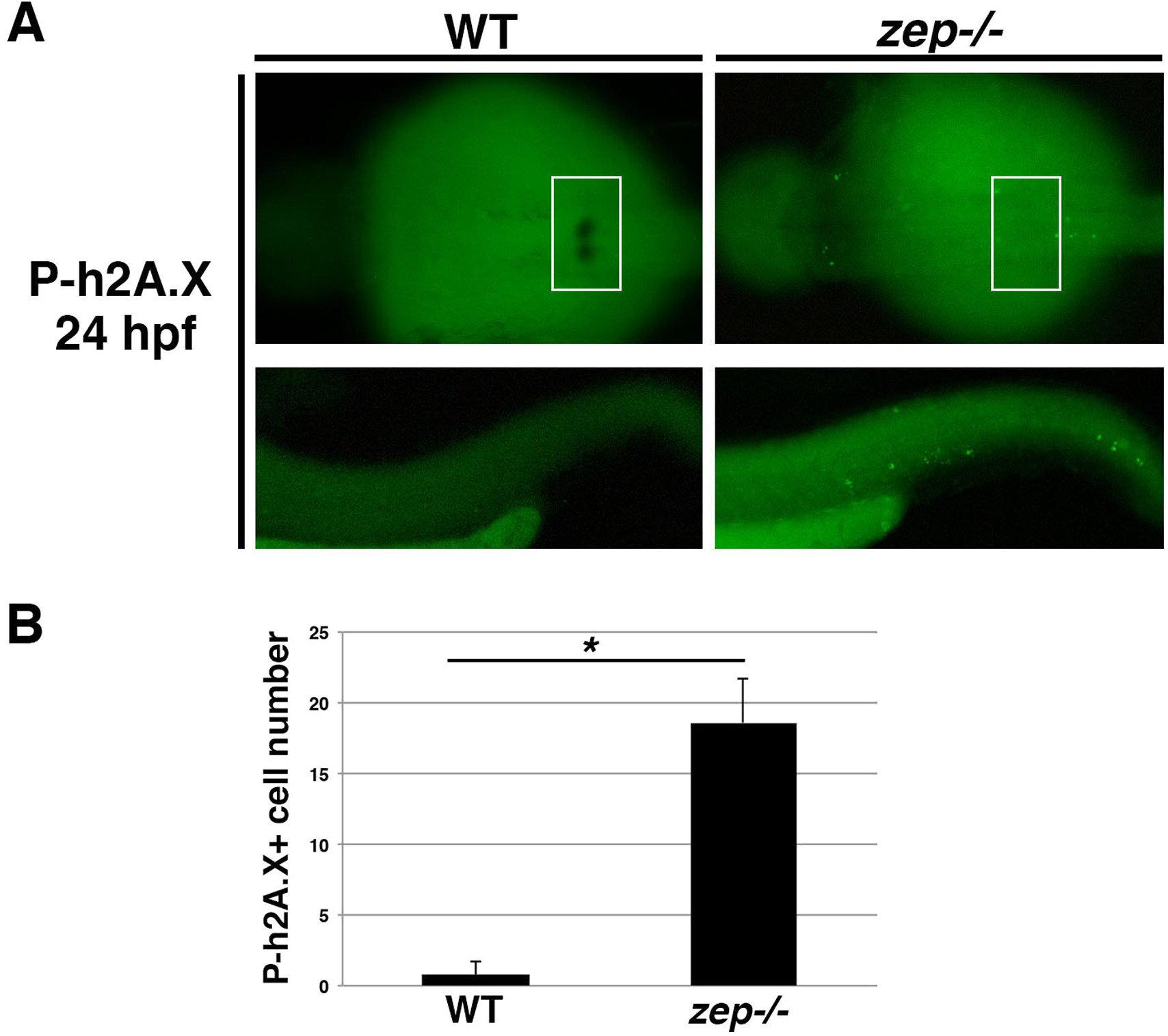
zepmutants exhibit elevated DNA damage. (A) WT and zep embryos were double labeled to detect podocytes by WISH for wt1b transcripts followed by whole mount immunohistochemistry to detect phosphorylated h2A.X (P-h2A.X), a marker of double strand breaks. Top panels show a dorsal view of the cervical region where podocytes develop, indicated by the white boxed region. Bottom panels show a lateral view of the tail, which was the area utilized for quantification. (B) Quantification of P-h2A.X+ cells in the tail region revealed a statistically significant increase in zep compared to WT. Asterisk (*) indicates p < 0.0001 using student T-test.
Reprinted from Developmental Biology, 428(1), Kroeger, P.T., Drummond, B.E., Miceli, R., McKernan, M., Gerlach, G.F., Marra, A.N., Fox, A., McCampbell, K.K., Leshchiner, I., Rodriguez-Mari, A., BreMiller, R., Thummel, R., Davidson, A.J., Postlethwait, J., Goessling, W., Wingert, R.A., The zebrafish kidney mutant zeppelin reveals that brca2/fancd1 is essential for pronephros development, 148-163, Copyright (2017) with permission from Elsevier. Full text @ Dev. Biol.