
Image
|
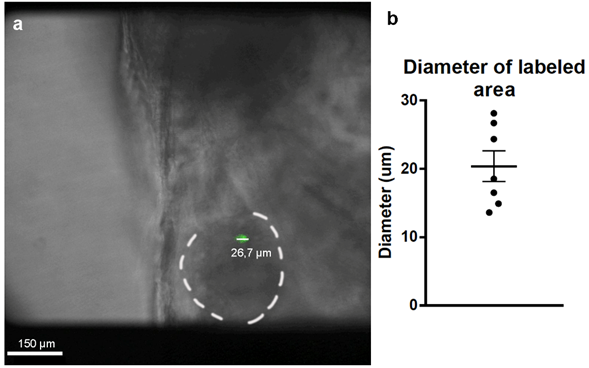
Figure Caption
Fig. S2
Diameters of the labeled areas in 4dpf zebrafish hearts. (a) Images of the labeled hearts were taken at 4 dpf and the diameters or the GFP-labeled areas were measured using ImageJ. (b) Measured diameters indicated that these labeled areas corresponded to one or two cardiomyocytes (n=7). Mean ± SEM.
Acknowledgments
This image is the copyrighted work of the attributed author or publisher, and
ZFIN has permission only to display this image to its users.
Additional permissions should be obtained from the applicable author or publisher of the image.
Full text @ Open Biol.