
|
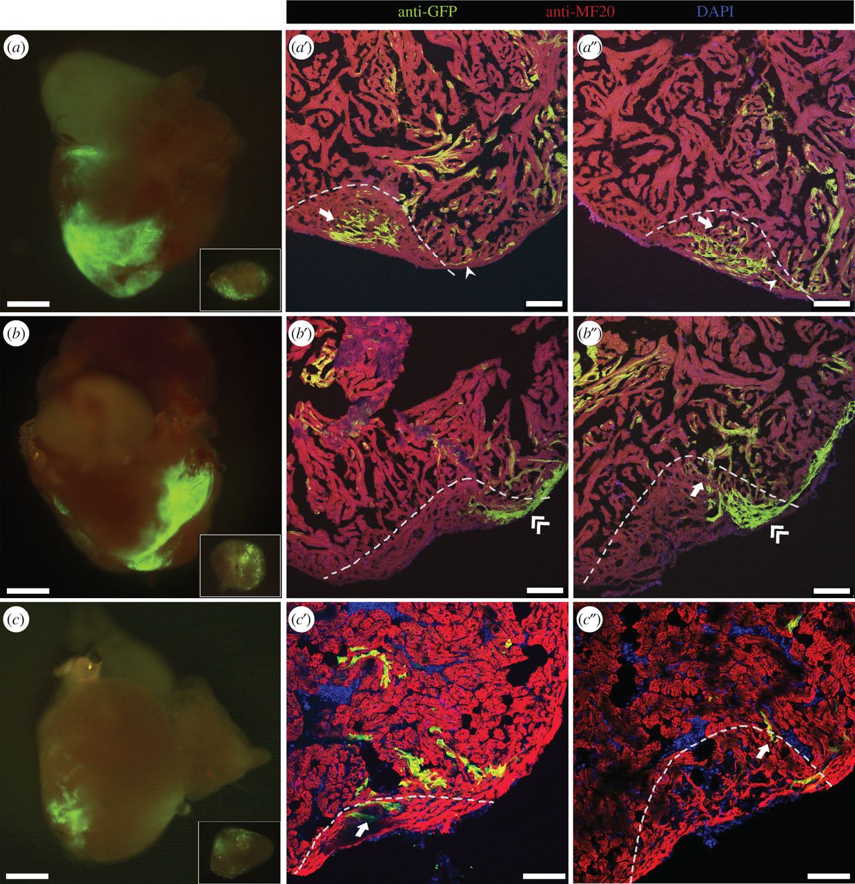
Fig. 3
Cell fate of labelled CMs during regeneration. Three-month-old adult zebrafish labelled at 2 dpf were subjected to ventricular amputation, and after 30 dpa the hearts were collected. (a–c) Epifluorescence images of representative hearts where the amputation plane passed through GFP-positive areas. The excised portion of tissue is shown in inset. (a′–c″) Collected hearts were sectioned, and processed for immunofluorescence with antibodies against MF20 and GFP. (a′,a″) Different sections of the regenerated myocardium of the heart shown in (a), where primordial (arrowhead) and trabecular (arrow) labelled CMs were observed. (b′,b″) Different sections of the regenerated myocardium of the heart shown in (b), where trabecular (arrow) and cortical (double-arrowhead) GFP-positive CMs were detected. (c′,c″) Different sections of the regenerated myocardium of the heart shown in (c), where trabecular (arrow) GFP-positive CMs were detected. The amputation plane is indicated by dashed lines. Nuclei were counterstained with DAPI. Scale bars: 500 µm in whole-mount hearts; 100 µm in sections.