
Fig. 4
- ID
- ZDB-IMAGE-170621-10
- Publication
- Solek et al., 2017 - Lineage tracing of dlx1a/2a and dlx5a/6a expressing cells in the developing zebrafish brain
- All Figures
- Figures for Solek et al., 2017
|
Fig. 4
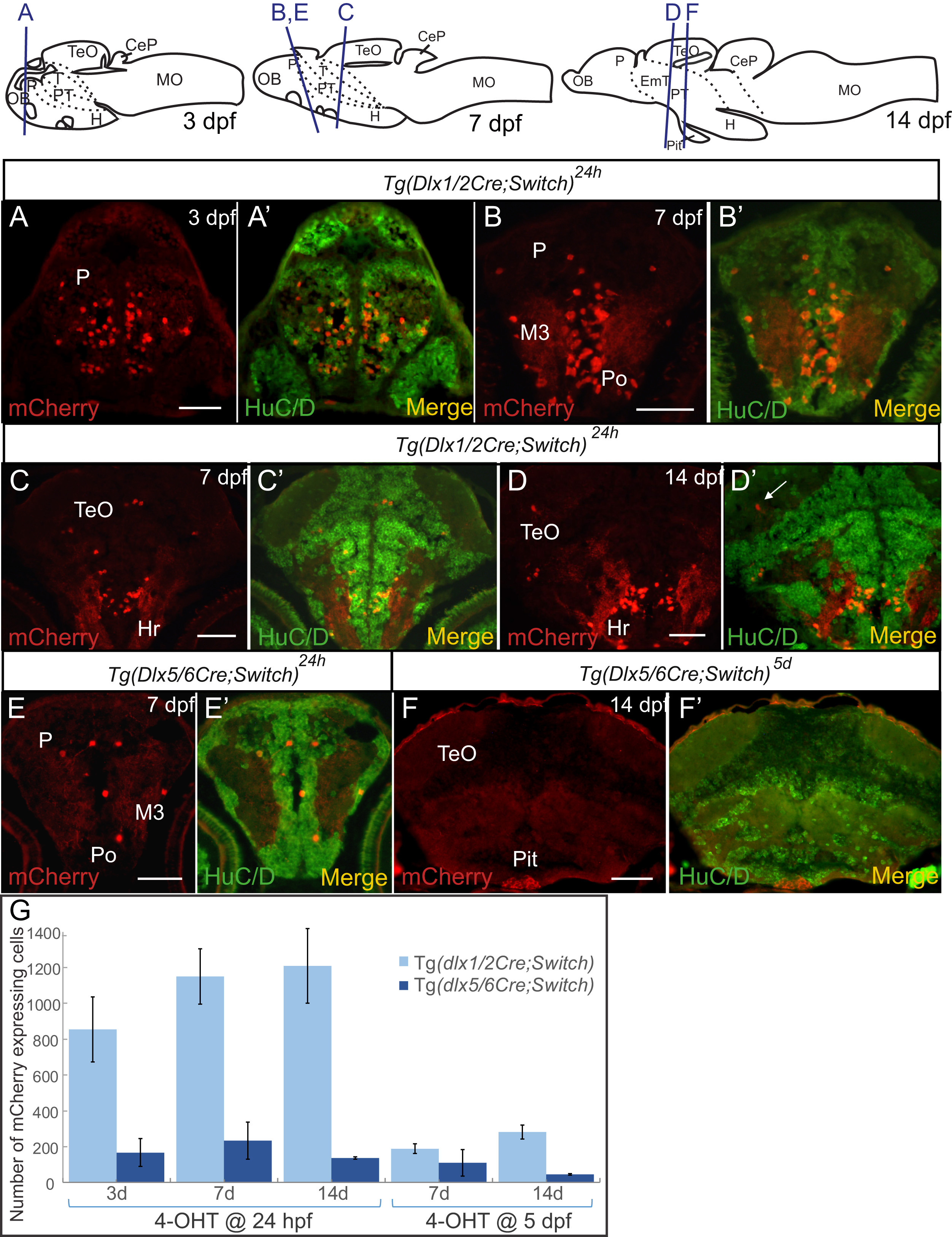
Colocalization of mCherry cells with the HuC/D neuronal marker in embryos and larvae. (A-F) Double immunohistochemical labeling of cryosections of embryos and larvae. Examples of staining of 3 dpf embryos (A), 7 dpf larvae (B, C, E) and 14 dpf larvae (D, F) are shown with relevant brain regions indicated. Each panel has mCherry staining alone (red, left) and merged image with HuC/D staining (green, right). A cell that expresses mCherry but not HuC/D in the neuropil is indicated by a white arrow in (D'). The location of each section is depicted in the cartoons at the top. (G) Number of mCherry expressing cells detected by immunohistochemistry in cryosections of zebrafish larvae from Tg(dlx1/2Cre;Switch) (light blue) and Tg(dlx5/6Cre;Switch) (dark blue) induced with 4-OHT at 24 hpf (left) and 5 dpf (right) in 3 dpf, 7 dpf and 14 dpf larvae. See Table S1 for sample sizes. Error bars: SEM. Scale bar: 100 µm.
Reprinted from Developmental Biology, 427(1), Solek, C.M., Feng, S., Perin, S., Weinschutz Mendes, H.C., Ekker, M., Lineage tracing of dlx1a/2a and dlx5a/6a expressing cells in the developing zebrafish brain, 131-147, Copyright (2017) with permission from Elsevier. Full text @ Dev. Biol.