
Image
|
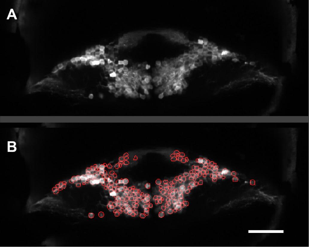
Figure Caption
Fig. S2
Automated segmentation of granule cell somata from whole-brain imaging data. Related to Figure 2.
A) Anatomical image of one imaging plane through the granule cell layer. B) Same plane with automated anatomical segmentation of granule cell somata indicated by red circles. In this plane, 252 cells were segmented. Scale bar = 50 microns.
Acknowledgments
This image is the copyrighted work of the attributed author or publisher, and
ZFIN has permission only to display this image to its users.
Additional permissions should be obtained from the applicable author or publisher of the image.
Full text @ Curr. Biol.