
|
Fig. S1
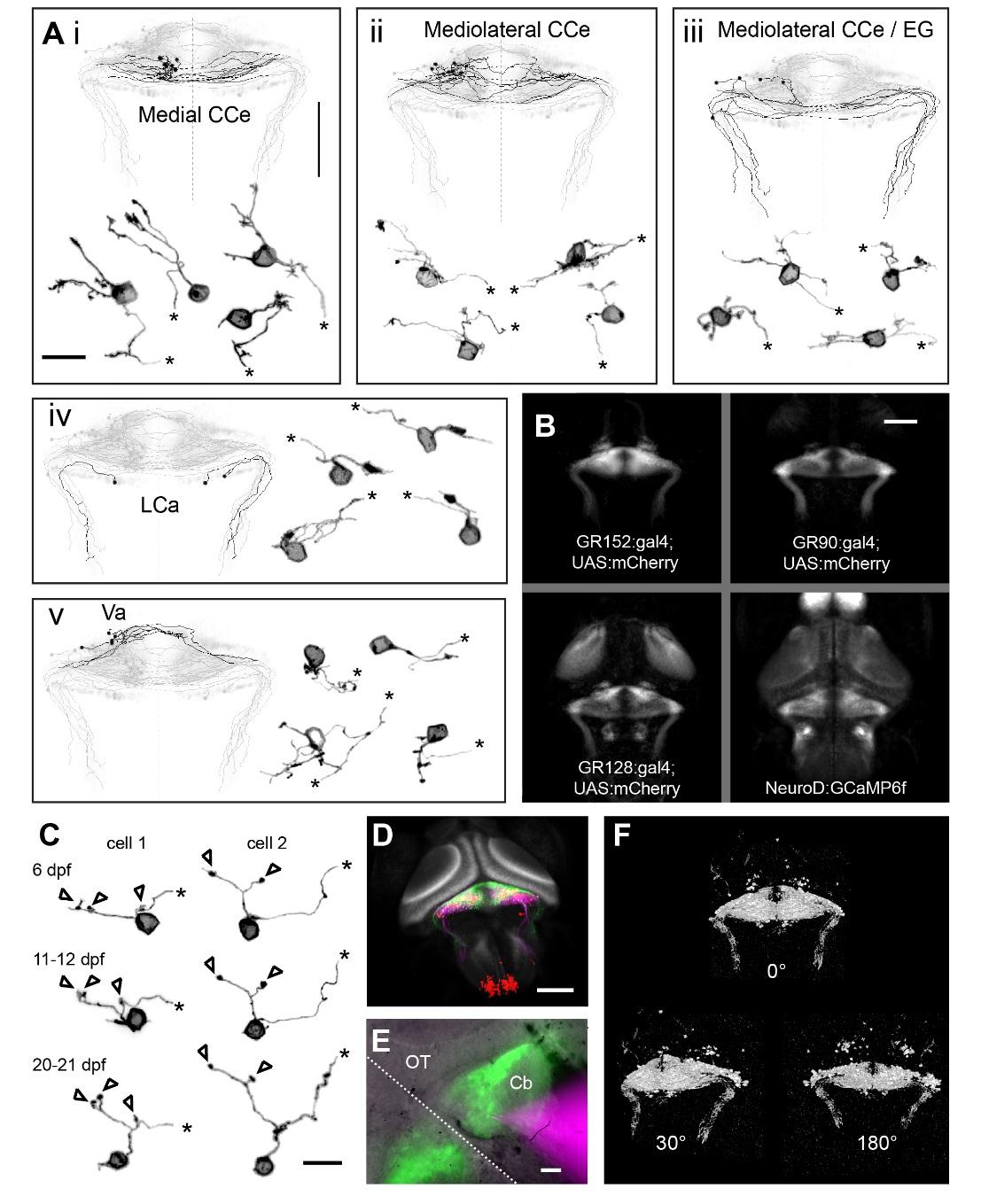
Detailed morphology of individual granule cells and transgenic populations. Related to Figure 1.
Ai-v) Example additional dendritic morphologies of twenty granule cells are grouped by somata location and axonal projection (refer to Figure 1 for information on abbreviated anatomical regions). Truncated parallel fibers in the detailed morphology are indicated by asterisks. Scale bar = 10 microns. The traced parallel fiber projections (shown in black) for these cells and others with somata in the same region are morphed to a common reference anatomy (shown in grey). Scale bar = 100 microns for the parallel fiber overviews. B) Z-projections of confocal stacks from transgenic lines labeling the granule cells used in our experiments. Morphologies were obtained from (clockwise, starting at upper left) N= 6, 5, 3, and 2 fish. Scale bar = 100 microns. C) Dendritic morphology of two granule cells in the CCe at three developmental time points showing the stability of dendritic claw number and overall morphology. Scale bar = 10 microns. D) Tilted view showing pan-neuronal GCaMP6f in the zebrafish brain in grey (the moon-shaped optic tecta are very salient). The cerebellum has a stereotypical tri-layered configuration where the granule cell layer (green) sends parallel fibers to the molecular layer, where they contact the dendrites of the Purkinje cells (magenta). Inferior olivary neurons (red) also provide inputs to the Purkinje cells. Scale bar = 100 microns. E) Composite whole-field image from a cell-attached electrophysiological recording of a granule cell. A large granule cell population is labelled by GR90:gal4;UAS:GFP (in green), the pipette (with intracellular solution containing sulforhodamine dye) is labelled in magenta, and the bright-field anatomy is shown in grey. Scale bar = 20 microns. F) Whole-brain imaging of fish expressing GCaMP6s in granule cells provided datasets such as the one shown here that can be easily segmented functionally by using local fluorescence correlations. All the significantly active voxels for one experiment are shown here at three different roll angles: 0 degrees (from above) showing parallel fibers, 30 degrees and 180 degrees (from below) showing granule cell somata (see also Movie S1). Scale same as for (B).