
|
Fig. 1
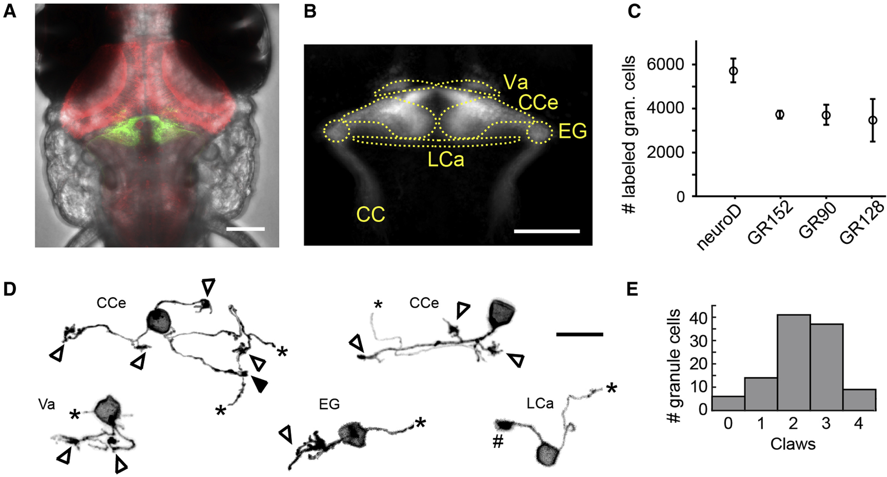
Characterization of the Larval Zebrafish Cerebellum and Its Granule Cell Population
(A) Composite confocal stack showing the head of a 7-dpf transgenic larval zebrafish in bright-field expressing GCaMP6f pan-neuronally (in red) and mCherry in a large population of granule cells (in green). The scale bar represents 100 μm.
(B) Schematic showing the anatomical lobes of the zebrafish cerebellum. CC, crista cerebellaris; CCe, corpus cerebella; EG, eminentia granularis; LCa, lobus caudalis cerebelli; Va, valvula cerebelli. The scale bar represents 100 μm.
(C) Quantification of the number of granule cells labeled in each of the four transgenic zebrafish lines used in this study (n = 3, 6, 5, and 2 fish, respectively). Data are represented as mean ± SEM.
(D) Example labeled granule cells from different regions of the cerebellum with zero, one, two, three, or four dendritic claws. Open arrowheads indicate dendritic claws, black arrowheads indicate dendritic branches without claws, pound indicates putative growth cone, and the truncated parallel fiber axons for all cells are marked with asterisks. The scale bar represents 10 μm. See Figure S1 for additional morphology of granule cells.
See also Figure S1.