
Fig. 3
- ID
- ZDB-IMAGE-170607-7
- Antibodies
- Publication
- Kelu et al., 2017 - Ca2+ release via two-pore channel type 2 (TPC2) is required for slow muscle cell myofibrillogenesis and myotomal patterning in intact zebrafish embryos.
- All Figures
- Figures for Kelu et al., 2017
|
Fig. 3
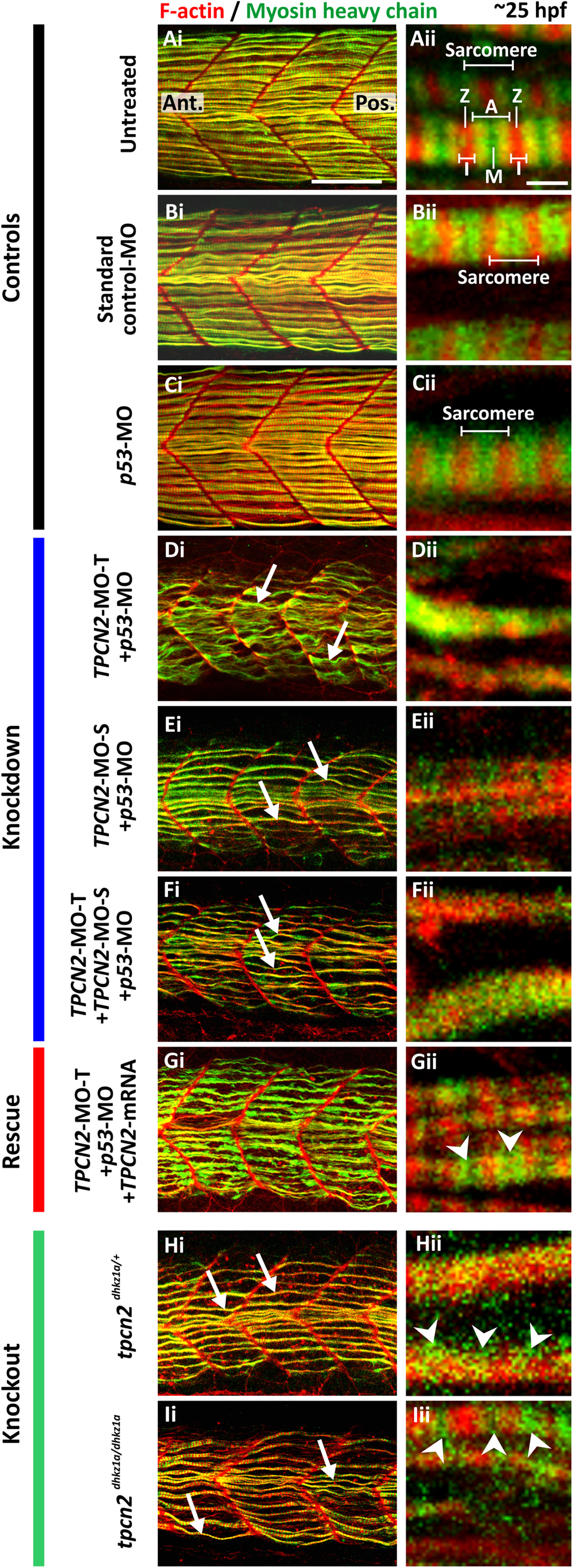
Effect of MO-based knockdown (without and with mRNA rescue) and CRISPR/Cas9-knockout of TPC2 on the organization of the trunk musculature and the formation of the sarcomeres. Embryos were (A) untreated or (B-G) injected with: (B) ~5 ng standard control-MO; (C) ~7.5 ng p53-MO; (D) ~2.5 ng TPCN2-MO-T with p53-MO; (E) ~5 ng TPCN2-MO-S with p53-MO; (F) ~1.3 ng TPCN2-MO-T+~2.5 ng TPCN2-MO-S with p53-MO; or (G) ~2.5 ng TPCN2-MO-T with p53-MO and ~50 pg tpcn2 mRNA. In addition, representative (H) heterozygous and (I) homozygous tpcn2 mutants are shown. All embryos were fixed at ~25 hpf and dual-labeled with phalloidin and the F59 antibody, to visualize F-actin (in red) and myosin heavy chain (in green) in the trunk musculature, respectively. Panels show a series of optical sections projected as single images at (Ai-Ii) low and (Aii-Iii) higher magnification when the red and green channels are merged; overlapping regions are shown in yellow. The higher magnification images of the SMC myofibers, reveal the presence or absence of the sarcomeric banding pattern of the F-actin and myosin heavy chain. The white arrows in panels Di-Fi, Hi-Ii show examples of elongated (flexuous) SMC myofibers; ‘A’, ‘I’, ‘M’ and ‘Z’ in panel Aii indicate the A-band, I-band, M-line and Z-line of a sarcomere respectively; and the white arrowheads in panels Gii-Iii indicate the appearance of some banding but clear sarcomeres are not obvious. Ant. and Pos. in panel Ai are anterior and posterior, respectively. Scale bars, 50 µm in panels Ai-Ii; and 2 µm, in panels Aii-Iii.
Reprinted from Developmental Biology, 425(2), Kelu, J.J., Webb, S.E., Parrington, J., Galione, A., Miller, A.L., Ca2+ release via two-pore channel type 2 (TPC2) is required for slow muscle cell myofibrillogenesis and myotomal patterning in intact zebrafish embryos., 109-129, Copyright (2017) with permission from Elsevier. Full text @ Dev. Biol.