IMAGE

Fig. 2

ID
ZDB-IMAGE-170606-15
Genes
Source
Figures for Yoo et al., 2017
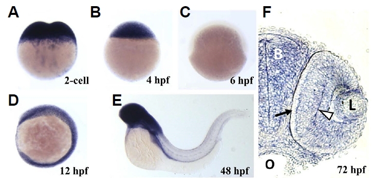
Image
Figure Caption

Fig. 2

Spatiotemporal expression of ranbp9 mRNA during embryonic development.

Whole-mount in situ hybridization was performed with DIG-labeled antisense RNA probe (hpf, hours after fertilization). (A) At 2-cell stage, maternal transcripts were detected in high density. (B) At dome stage (4 hpf), high level of transcription persisted. (C) At the beginning of gastrulation, shield stage (6 hpf), ranbp9 mRNA was hardly detected. (D) At 6-somite stage (12 hpf), ranbp9 was seen expressed ubiquitously. (E) At 48 hpf, ranbp9 expression was mainly observed in the head region. (F) Section of head region at 72 hpf, showing ranbp9 expression in the brain (B) and eye (L, lens). Its expression is more prominent in forming layers (arrow head) of the retina, compared to the periphery (arrow). O, oral cavity.

Figure Data
Acknowledgments
This image is the copyrighted work of the attributed author or publisher, and ZFIN has permission only to display this image to its users. Additional permissions should be obtained from the applicable author or publisher of the image. Full text @ Mol. Cells