
Fig. 7
- ID
- ZDB-IMAGE-170512-30
- Genes
- Antibodies
- Publication
- Berberoglu et al., 2017 - Satellite-like cells contribute to pax7-dependent skeletal muscle repair in adult zebrafish
- All Figures
- Figures for Berberoglu et al., 2017
|
Fig. 7
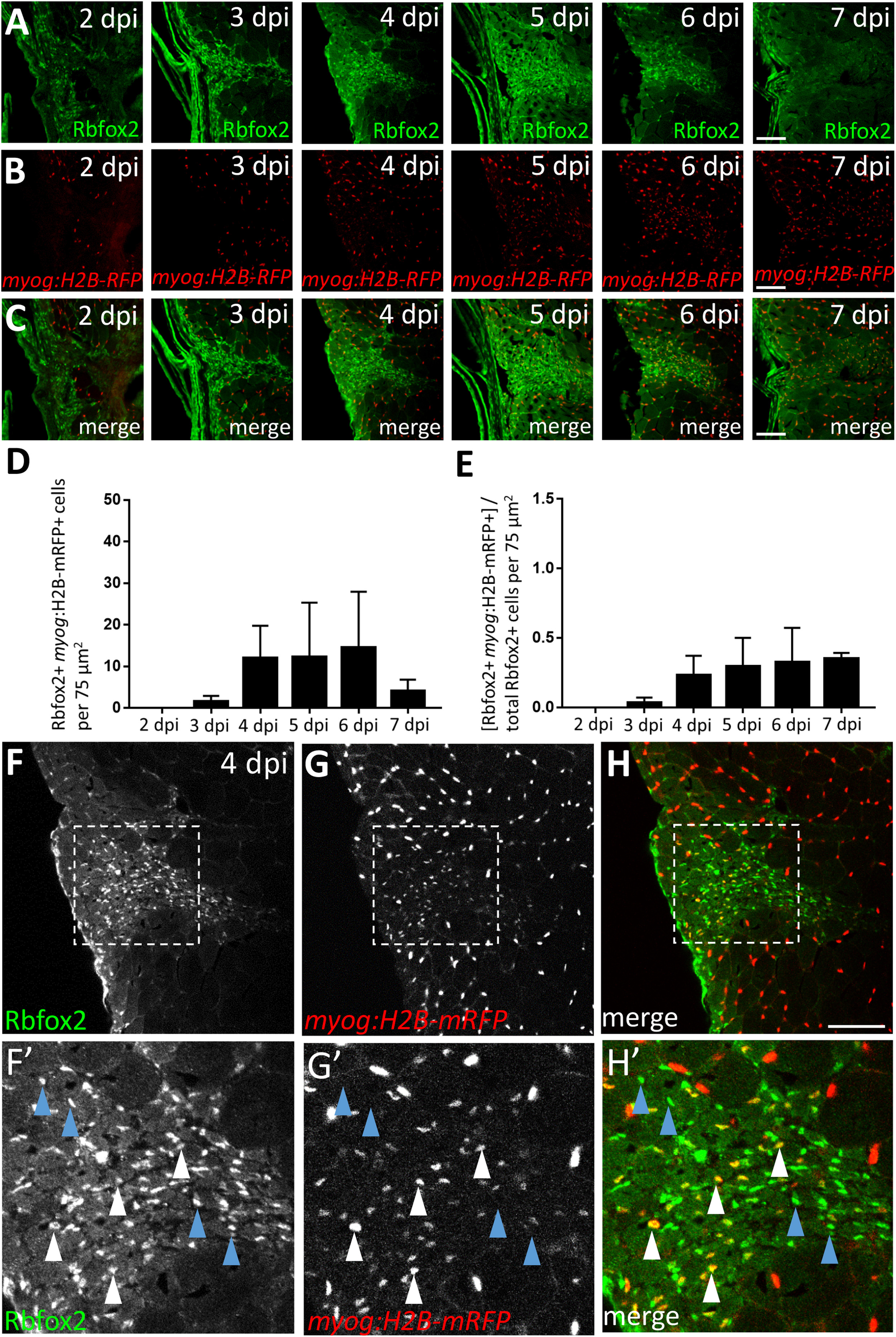
A subset of Rbfox2-expressing cells are myog:H2B-mRFP-positive during skeletal muscle repair. (A) Injury time-course experiment performed in myog:H2B-mRFP transgenic line showing Rbfox2 expression (on alternating sections with those described in Fig. 6). (B) As shown in Fig. 6B, myog:H2B-mRFP expression is readily observed within the injury site by 4 dpi. (C) Overlay of Rbfox2 and myog:H2B-mRFP expression. (D) Total number of Rbfox2, myog:H2B-mRFP double-positive cells at 2–7 dpi. (E) Ratio of Rbfox2-positive cells that are myog:H2B-mRFP-positive at 2–7 dpi. One-way ANOVA followed by Tukey's multiple comparisons test was performed for all of the above statistical analyses; differences among time-points are not statistically significant. (F-H) Higher magnification view of 4 dpi injury site shown in A-C. (F’-H’) Area shown is the boxed region in F-H that highlights examples of Rbfox2-expressing cells that are myog:H2B-mRFP-positive (white arrowheads) and myog:H2B-mRFP-negative (blue arrowheads). Scale bar in all panels is 75 µm.
Reprinted from Developmental Biology, 424(2), Berberoglu, M.A., Gallagher, T.L., Morrow, Z.T., Talbot, J.C., Hromowyk, K.J., Tenente, I.M., Langenau, D.M., Amacher, S.L., Satellite-like cells contribute to pax7-dependent skeletal muscle repair in adult zebrafish, 162-180, Copyright (2017) with permission from Elsevier. Full text @ Dev. Biol.