
IMAGE
Fig. S1
- ID
- ZDB-IMAGE-170421-6
- Publication
- Goldman et al., 2017 - Resolving Heart Regeneration by Replacement Histone Profiling
- All Figures
- Figures for Goldman et al., 2017
Image
|
Figure Caption
Fig. S1
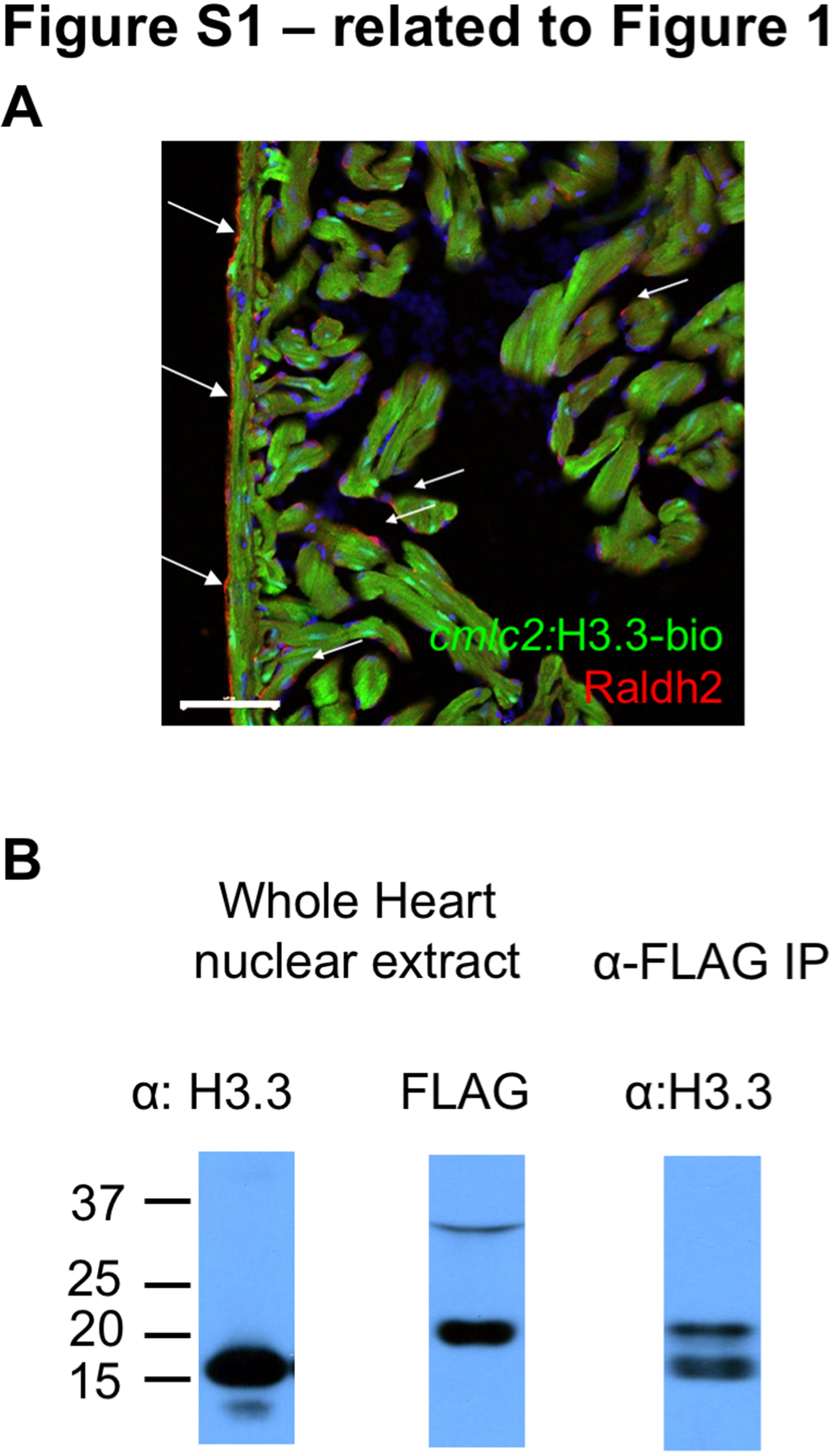
H3.3-bio is Present Sub-stoichiometrically to Endogenous H3.3 Protein (related to Figure 1)
(A) cmlc2:H3.3-bio does not drive expression (green) in epicardium and endocardium. Both non-muscle cell types are recognized by an antibody against Raldh2 (arrows; red). Scale bar, 50 μm.
(B) Endogenous H3.3 is expressed in excess of transgenic H3.3-bio, which is undetectable using anti-H3.3 (lane 1). However, H3.3-bio can be detected from whole hearts using anti-FLAG (lane 2). Immunoprecipitation of H3.3-bio chromatin using the FLAG antibody enriches both forms of H3.3 (lane 3).
Acknowledgments
This image is the copyrighted work of the attributed author or publisher, and
ZFIN has permission only to display this image to its users.
Additional permissions should be obtained from the applicable author or publisher of the image.
Reprinted from Developmental Cell, 40, Goldman, J.A., Kuzu, G., Lee, N., Karasik, J., Gemberling, M., Foglia, M.J., Karra, R., Dickson, A.L., Sun, F., Tolstorukov, M.Y., Poss, K.D., Resolving Heart Regeneration by Replacement Histone Profiling, 392-404.e5, Copyright (2017) with permission from Elsevier. Full text @ Dev. Cell