Fig. S1
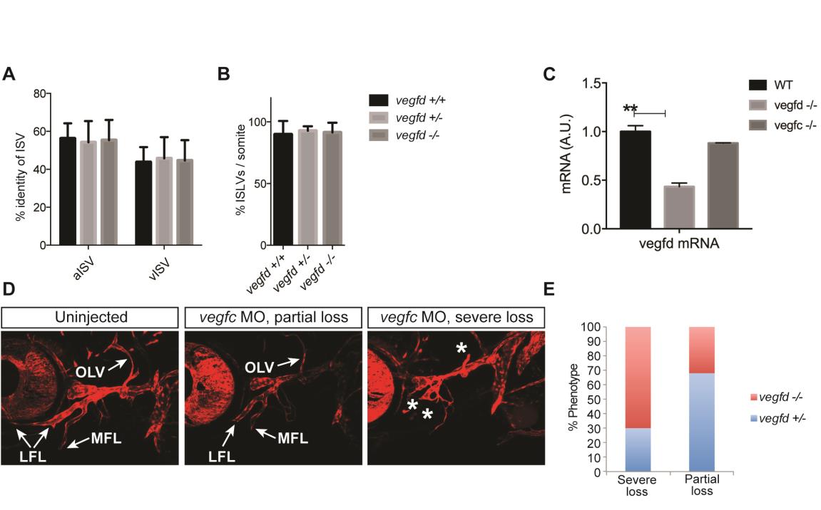
Characterisation and quantification of vessels in vegfduq9bh mutant embryos. (A) Quantification of the percentage of arterial intersegmental vessels (aISVs) and venous intersegmental vessels (vISVs) at 5dpf per somite across 10 somites anterior from the end of the yolk extension in vegfduq9bh heterozygous, mutant and wildtype embryos reveals no significant differences between genotypes. Error bars represent mean +/- sem; one way ANOVA analysis (n=6). (B) Quantification of the percentage of ISLVs at 5dpf per somite across 10 somites anterior from the end of the yolk extension in vegfduq9bh heterozygous, mutant and wildtype embryos reveals no significant differences between genotypes. Error bars represent mean +/- sem; one way ANOVA analysis (n=6). (C) Analysis of vegfd mRNA levels in embryos derived from an incross of vegfd adult mutants showing mutant embryos had approximately 50% of the transcript levels of wildtype embryos, suggesting non-sense mediated decay of mutant transcripts There was no change in vegfc mRNA levels in vegfduq9bh mutants indicating that compensation for the loss of vegfd does not occur in zebrafish. (D) Embryos from a vegfduq9bh heterozygote x vegfduq9bh homozygous mutant cross, injected with vegfc morpholino, were characterised for the loss of facial lymphatics as severe, moderate or none. (E) Subsequent genotyping found that the vegfduq9bh mutant embryos were enriched in the severe loss of facial lymphatic category.

