
Image
|
Figure Caption
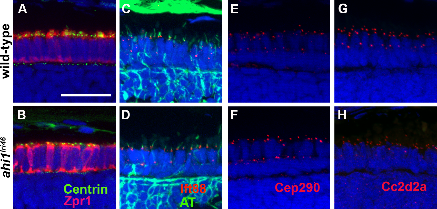
Fig. 9
ahi1 is not required for ciliogenesis or transition zone protein localization. (A, B) Tg(centrin:GFP) expression (green) localized to the apical inner segments in wild-type and ahi1lri46 mutants at 3 dpf. Zpr1 (red) was used to stain cone photoreceptors to visualize the apical boundary. (C, D) Five dpf retinas stained with acetylated tubulin (green) and Ift88 (red) showed ciliary localization of the IFT particle in wild-type and mutant animals. (E, H) The transition zone proteins Cep290 and Cc2d2a (red) exhibited punctate staining in wild-type and mutant animals. Scale bar: 20 μm.
Figure Data
Acknowledgments
This image is the copyrighted work of the attributed author or publisher, and
ZFIN has permission only to display this image to its users.
Additional permissions should be obtained from the applicable author or publisher of the image.
Full text @ Invest. Ophthalmol. Vis. Sci.