
|
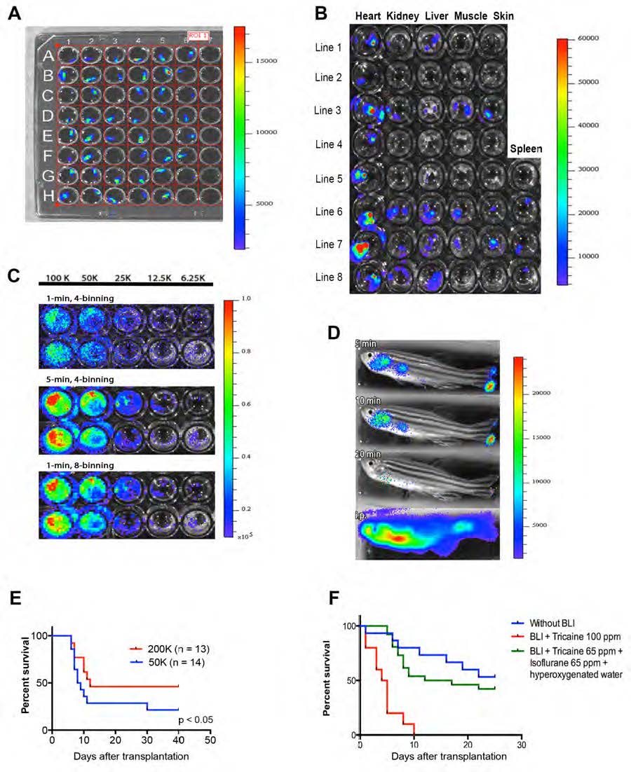
Fig. S1
Luciferase expression in ubi:luc zebrafish and modifications of BLI to detect WKM cells.
(A) Single cell stage zebrafish were injected with 100 ng/μL of the Tol2 ubi:luc construct. Founder ubi:luc zebrafish embryos (96 hours post fertilization) were placed in a 96-well plate after the addition of D-luciferin to the water .
(B) Organs dissected from F2 zebrafish and BLI performed after the addition of Dluciferin to the water (150 mg/mL) indicating BLI variability in several lines.
(C) Limiting dilution with WKM from ubi:luc zebrafish in a 96-well plate followed by the addition 150 μg/mL D-luciferin to the water. Acquisition time set at 1 minute and CCD resolution set at 4 pixel binning gave good signal sensitivity but high background scatter. When acquisition time was increased to 5 minutes (middle panel) or resolution set to 8 pixel binning (lower panel), both BLI sensitivity and localization improved without interference from background signal.
(D) Bathing versus intraperitoneal injection of D-luciferin in transplant recipients. Lethally irradiated wild-type zebrafish received 100,000 ubi:luc WKM cells and allowed to engraft for 28 days. Imaging was performed using a Xenogen IVIS50 (Stage A). Exposure time was set at 1 minute. Animals were immobilized using 0.005% tricaine in a 6-well plate filled with 1% agarose gel with a portion removed to isolate the animal. Bathing was performed by placing zebrafish in D-luciferin in fish water at 150 μg/mL for the indicated lengths of time (top three panels). Intraperitoneal injection (lower panel) of 75 μg D-luciferin was performed immediately prior to BLI imaging (exposure time was set at 1 minute as before).
(E) Short-term survival is increased in zebrafish receiving a higher cell dose after HCT. Recipients received 30 Gy, followed by HCT with 50,000 or 200,000 ubi:luc WKM cells. Animals were followed for 40 days. P-value from a Student’s t-test.
(F) Modification to anesthesia protocol. Wild-type recipients underwent HCT with 200,000 ubi:luc donor cells with BLI performed at 1 dpt and 5 dpt using the anesthesia as listed, n = 10/group.
All luminescence scale bars show radiance (p/sec/cm2/sr).