IMAGE

Fig. 1

ID
ZDB-IMAGE-170222-127
Source
Figures for Huemer et al., 2017
Image
Figure Caption

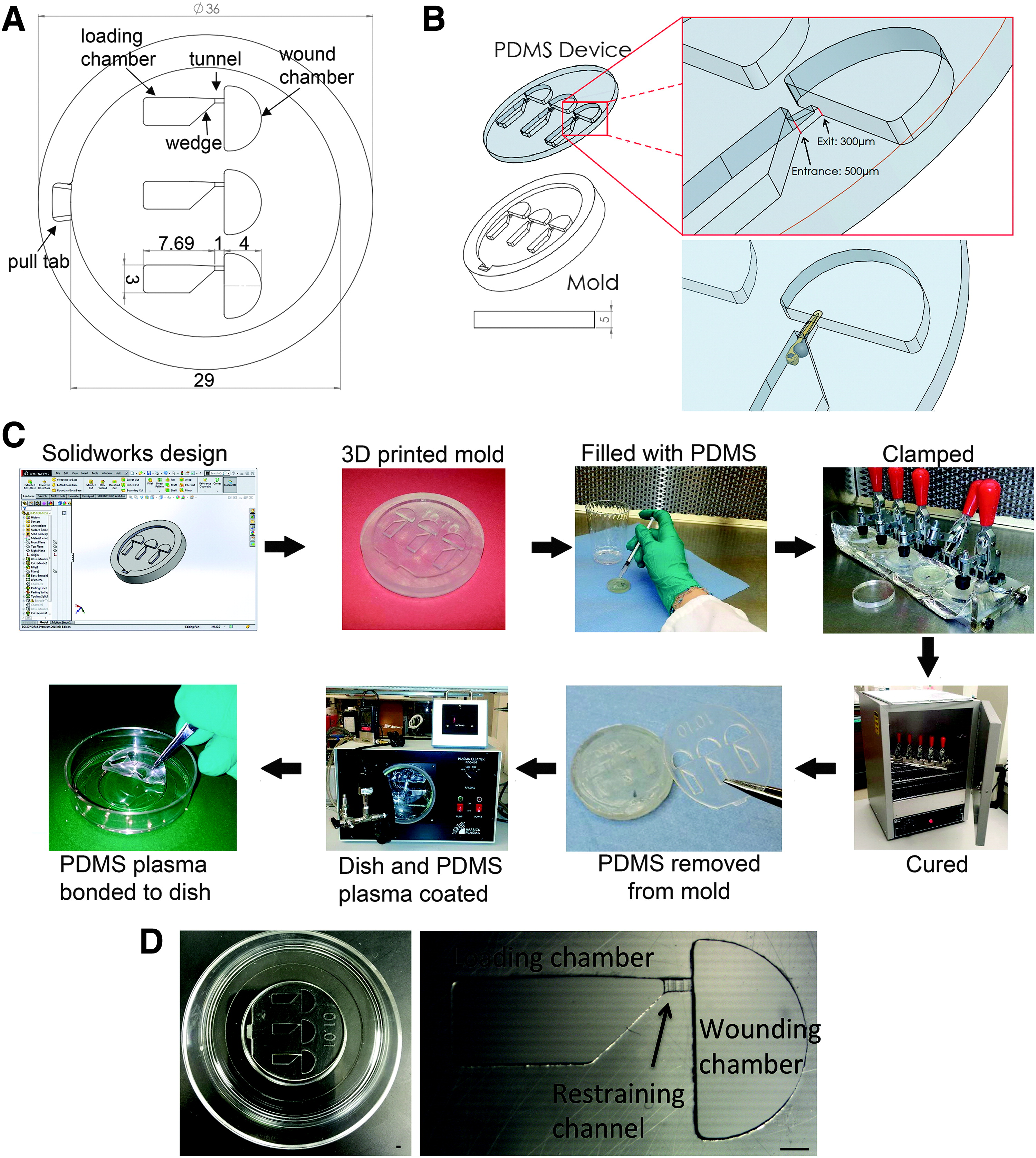
Fig. 1

(A) Labeled and dimensioned enface SolidWorks drawing of the zWEDGI mold. (B) SolidWorks isometric of mold and PDMS device. Upper expanded view details the structure of the restraining tunnel and lower view shows positioning of larva. All dimensions are in mm unless specifically noted. (C) Flow chart illustrating procedure for making zWEDGI. Details of the procedure are in the Materials and Methods section. (D) Finished PDMS device plasma bonded to glass bottom dish and a detailed view of a single channel with compartments labeled. Scale bar = 1 mm. PDMS, polydimethylsiloxane; zWEDGI, zebrafish Wounding and Entrapment Device for Growth and Imaging.

Acknowledgments
This image is the copyrighted work of the attributed author or publisher, and ZFIN has permission only to display this image to its users. Additional permissions should be obtained from the applicable author or publisher of the image. Full text @ Zebrafish