
|
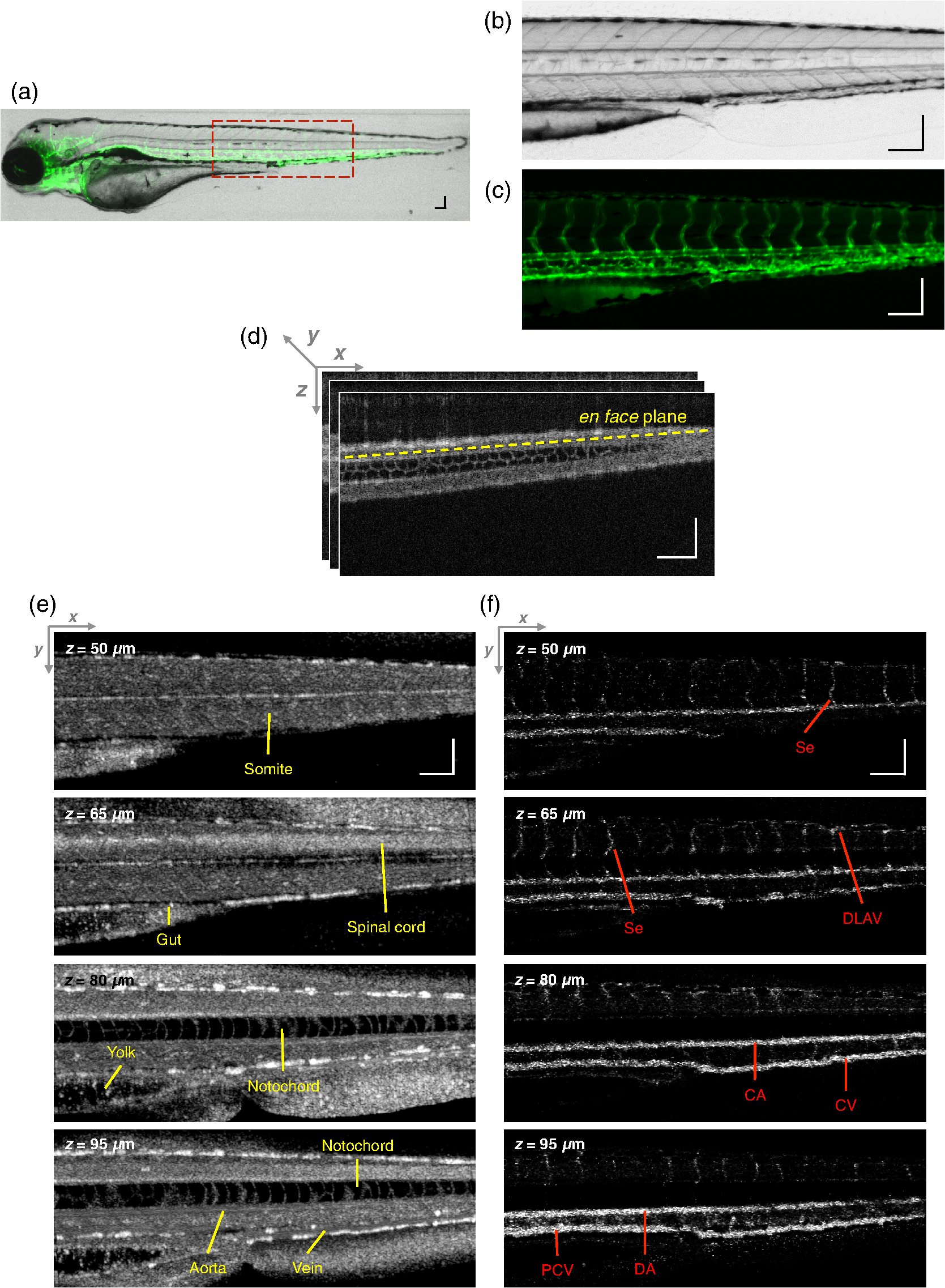
Fig. 5
Structural and vascular imaging of zebrafish embryos. (a) Composite wide-field microscopic image of a 3--dpf zebrafish embryo: Tg(kdrl:eGFP). The red dashed box marks the trunk area of interest for SD-OCM imaging. (b) Bright-field microscopic image of the structure of a 3-dpf zebrafish trunk. (c) Fluorescence microscopic image of the GFP-labeled vascular endothelium in the trunk. (d) OCM B-scan intensity images at roughly the same area. The yellow dashed line represents one example plane for creating an en face image. (e) En face OCM intensity images at various depth layers. (f) En face pvOCM images of the vasculature at various depth layers. In both (e) and (f), each en face image was created from average intensity projection of 5-μm depth and spanned an area of 1.20×0.45 mm2. The depth z was relative to the top surface of the zebrafish trunk. Scale bars: 100 μm.