
Fig. 3
- ID
- ZDB-IMAGE-161206-58
- Genes
- Publication
- Zada et al., 2016 - Pharmacological and BBB-targeted genetic therapies for thyroid hormone-dependent hypomyelination
- All Figures
- Figures for Zada et al., 2016
|
Fig. 3
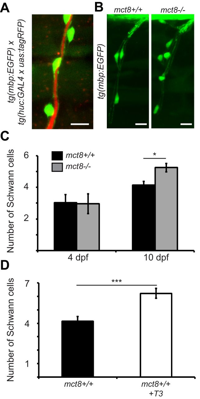
Increased number of Schwann cells in mct8−/− larvae during development. (A) Lateral view of a motor neuron in 10-dpf live tg(mbp:EGFP)/tg(huc:GAL4 x uas:tagRFP) larvae shows Schwann cell (green) ensheathment of motor neuron axons (red). (B) Representative lateral-view images of Schwann cell ensheathment of motor neuron axons in the SC of 10-dpf mct8+/+ and mct8−/− larvae. (C) Number of Schwann cells that myelinate the axons of the motor neurons of 4 (mct8+/+: n=9, mct8−/−: n=9)- and 10 (mct8+/+: n=26, mct8−/−: n=22)-dpf larvae. (D) Number of Schwann cells that myelinate the axons of the motor neurons of untreated and T3-treated mct8+/+ 10-dpf larvae (untreated: n=26, T3-treated: n=19). Values are represented as means±s.e.m. Statistical significance was determined by Student's t-test: two samples assuming unequal variance (*P<0.05, ***P<0.001). Scale bars: 10 µm.