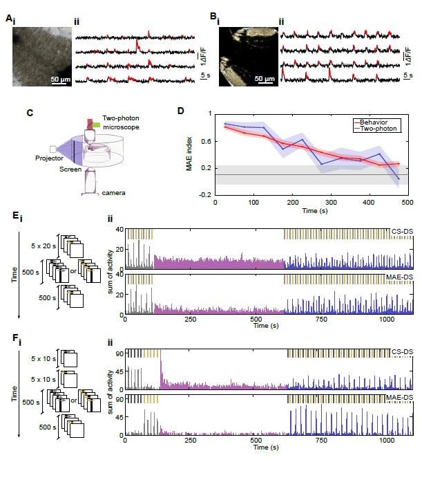
Fig. S2
Calcium-imaging-data-processing methods and experimental protocols (related to Figure 4).
(A) i. An optical section of the zebrafish larva’s optic tectum superimposed with the ROIs corresponding to each single neurons (yellow patches). ii. Examples of typical single-neuron DF/F traces (black) with significant fluorescence transients highlighted in red. Breaks in the traces depict discarded frames due to movement artifacts.
(B) i. An optical section of the optic tectum of an ath5:GCaMP3 zebrafish larva, where the tectal neuropil can be clearly observed. The SROIs are superimposed to the image (yellow patches). ii. Examples of typical single-SROIs DF/F traces (black) with significant fluorescence transients highlighted in red. Breaks in the traces depict discarded frames due to movement artifacts.
(C) Experimental setup: Scheme of the two-photon system for simultaneously monitoring eye movements and presenting visual stimuli.
(D) MAE index as a function of time after CS under the two photon microscope. Blue curve: MAE index as a function of time under a two-photon microscope. CS consisted in repetitive moving bars for 500 s. Red curve: MAE index as a function of time as showed in Fig. 1F. CS consisted in whole field grating for 500 s. Gray line represents the control index for the two-photon experiments. Nonsignificant differences where found between the two conditions. Error bars: s.e. For repetitive moving bars, n=20 (trials), from 10 larvae. For whole field grating, n=15 (trials), from 15 larvae.
(E) i. Experimental paradigm for monitoring adaptation of RGC projections in paralyzed larvae. ii. The sum of the relative change in fluorescence intensity (DF/F) of direction selective SROIs as a function of time. Top panel, CS direction selective SROIs. Bottom panel, MAE direction selective SROIs. The color bars represent the different stimulation blocks of the experimental paradigm: gray for pre-CS control, magenta for CS and blue post-CS control period. Top bars: depict the presentation period of each moving bar (gray: CS direction, yellow: MAE direction).
(F) As in E, but for tectal neuron recordings.

