
Fig. S1
- ID
- ZDB-IMAGE-161103-13
- Publication
- Sicca et al., 2016 - Gain-of-function defects of astrocytic Kir4.1 channels in children with autism spectrum disorders and epilepsy
- All Figures
- Figures for Sicca et al., 2016
|
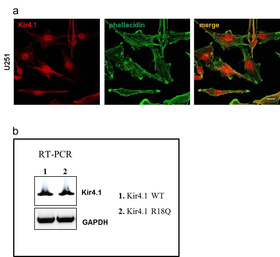
Fig. S1
WT and mutated Kir4.1 expression and distribution in U251 astrocytoma cells. (a) Immunofluorescence stainings of U251 astrocytoma cells with anti-Kir4.1 pAb (red) and FITC-conjugated phallacidin (green) to stain actin filaments show that the endogenous Kir4.1 channels are mainly distributed in cytoplasmic perinuclear area and scarcely at plasma membrane. Scale bars: 10 µm. (b) RT-PCR analysis using primers to detect specifically recombinant Kir4.1 mRNA expression (Forward: Xpress epitope/pcDNA 3.1/His, Life technologies; Kir4.1 Reverse: TCAGACATTGCTGATGCGCAC) in stably infected U251 cells reveals no differences between WT (1) and R18Q Kir4.1 (2) expressing cells. GAPDH housekeeping gene normalizes the amount of template used.