
|
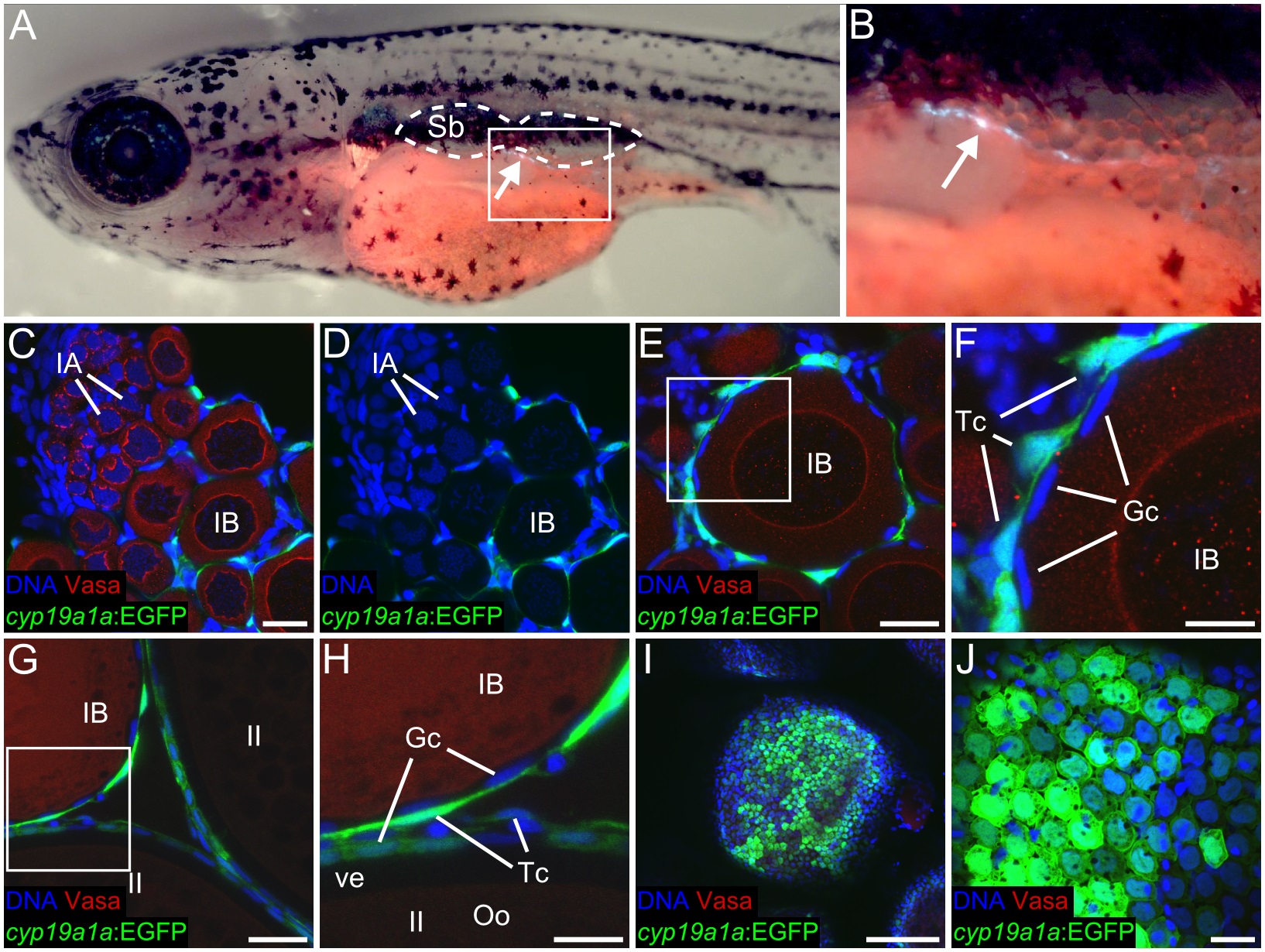
Fig. 3
(A-C) cyp19a1a:EGFP expression in the gonad at 16 dpf. (A) Anterior body of 16 dpf TgBAC(cyp19a1a:EGFP) fish showing GFP-expressing somatic gonad cells (arrow) located below the swim bladder (outlined). (B) Magnified view of the region boxed in (A). (C-F) cyp19a1a:EGFP is expressed in theca cells that surround stage IB and later stage oocytes (E-H), but not IA oocytes or premeiotic germ cells (C, D), as observed here in a 40 dpf ovary. (F) Magnified view of the region boxed in (E). (G-J) cyp19a1a: EGFP is expressed in both theca and granulosa cells that surround stage II and later stage oocytes, as observed here in a 60 dpf ovary. (H) magnified view of region boxed in (G). (I, J) low and high magnification views of cyp19a1a:EGFP expression in granulosa cells that are on the surface of a stage III oocyte. In all images, cyp19a1a:EGFP is green, Vasa protein is red and DNA is blue. Sb, swim bladder; IA, stage IA oocytes; IB, stage IB oocyte; II, stage II oocyte; Tc, theca cell; Gc, granulosa cell; Oo, oocyte; ve, vitelline envelope. Scale bars: 20 µm in C (for C, D, E, G, J), 10 µm in F (for F and H), 150 µm (I).