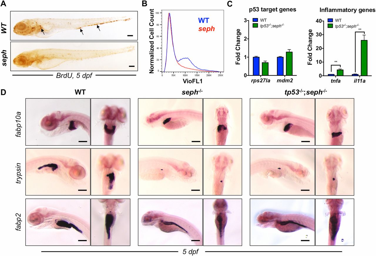
Fig. S4
p53 activation and cell cycle arrest contribute to the SepH-deficient phenotype. (A) Whole-mount immunohistochemical analysis of BrdU incorporation in WT and seph mutants at 5 dpf. Arrows point to clusters of BrdU-positive cells. (Scale bar: 200 µm.) (B) FACS analysis of the cell cycle in WT (blue) and seph mutants (red) at 5 dpf as determined by Hoechst staining (VioFL1). (C) qPCR expression analysis of specific genes associated with p53 activation (rps27l and mdm2) and an inflammatory response (tnfa and il11a) in WT and tp53-/-;seph-/- mutant larvae at 5 dpf. n = 3. **P < 0.01. (D) WISH analysis of fabp10a (liver), trypsin (pancreas), and fabp2 (intestine) expression in WT, seph-/-, and tp53-/-;seph-/- mutant larvae at 5 dpf. (Scale bar: 200 µm.)

