
IMAGE
Fig. 5
- ID
- ZDB-IMAGE-161005-19
- Publication
- Weinstein et al., 1996 - Hematopoietic mutations in the zebrafish
- All Figures
- Figures for Weinstein et al., 1996
Image
|
Figure Caption
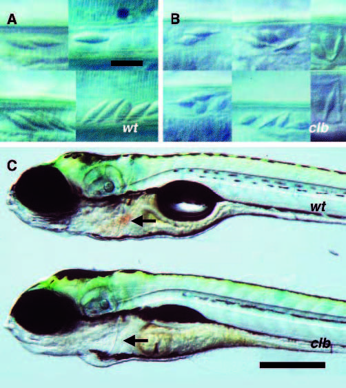
Fig. 5
Defects in erythrocyte morphology and hemoglobin accumulation in clb mutants. Embryos were obtained from a cross between two clbm525 heterozygotes. (A,B) DIC photomicroscopy of blood cells in the caudal arteries of 5 dpf phenotypically wild-type (A) and mutant (B) embryos. (C) Photomicrograph of anterior portions of live 3-day old phenotypically wild-type (top) and mutant (bottom) embryos. The heart appears visibly red (arrow) in the wildtype but not the mutant embryo (arrows). Scale bars, 10 µm (A,B), 400 µm (C).
Figure Data
Acknowledgments
This image is the copyrighted work of the attributed author or publisher, and
ZFIN has permission only to display this image to its users.
Additional permissions should be obtained from the applicable author or publisher of the image.
Full text @ Development