
Image
|
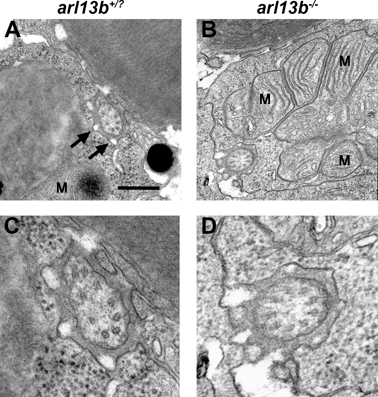
Figure Caption
Fig. 2
Ultrastructural analysis of photoreceptor connecting cilia by transmission electron microscopy. (A, B). Horizontal sections through photoreceptors of wild-type and arl13b-/- larvae at 5 dpf show the connecting cilia in the vicinity of the mitochondria (M). Calyceal processes surround the cilium (arrows). (C, D) Higher magnification images revealed no obvious defects in microtubule organization in arl13b-/- mutants. Scale bars: 0.5 µm (A, B) and 0.2 µm (C, D).
Figure Data
Acknowledgments
This image is the copyrighted work of the attributed author or publisher, and
ZFIN has permission only to display this image to its users.
Additional permissions should be obtained from the applicable author or publisher of the image.
Full text @ Invest. Ophthalmol. Vis. Sci.