
Fig. 7
- ID
- ZDB-IMAGE-160928-15
- Genes
- Publication
- Sarmah et al., 2016 - Embryonic Ethanol Exposure Dysregulates BMP and Notch Signaling, Leading to Persistent Atrio-Ventricular Valve Defects in Zebrafish
- All Figures
- Figures for Sarmah et al., 2016
|
Fig. 7
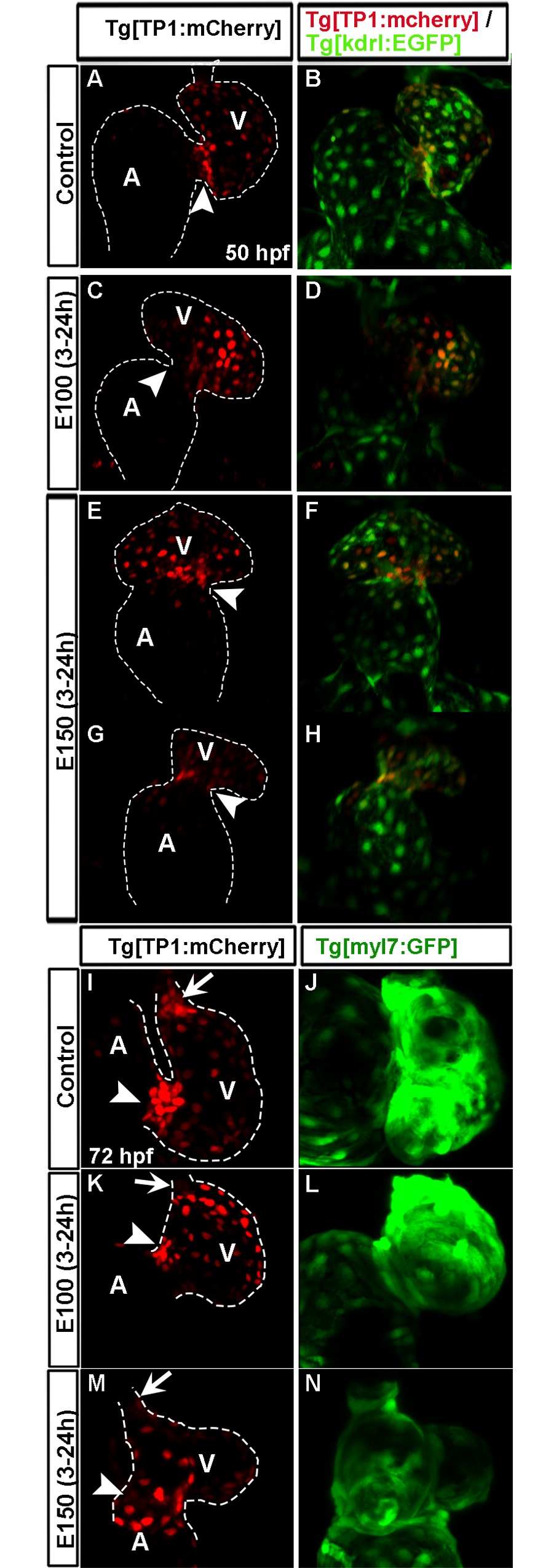
High Notch activity was not restricted at the AVC endocardial cells in ethanol exposed embryos.
(A-H) Tg(TP1:mCherry);Tg(kdrl:EGFP) double transgenic embryos showed compact cluster of strong mCherry labeled cells at the AVC and weak mCherry labeled cells in the ventricular endocardial lining in control embryo at 50 hpf (A, B); ethanol treated embryos had a mix of strong and weak mCherry labeled cells in the endocardium and no cluster of strong mCherry labeled cell at the AVC (C-H); severely defective embryo exhibited weaker mCherry labeling relative to the control (G, H). (I-N) Tg(TP1:mCherry);Tg(myl7:GFP) double transgenic embryos showed compact clusters of strong mCherry labeled cells at the AVC and OFT at 72 hpf in control (I) and no clustering in ethanol treated embryos (K, M); green fluorescence in Tg(TP1:mCherry);Tg(myl7:GFP) embryos showed myocardium at 72 hpf (J, L, N). A: Atrium, V: Ventricle, Arrowheads: AVC, Arrows: OFT.