
Fig. S1
- ID
- ZDB-IMAGE-160923-6
- Genes
- Publication
- Qiu et al., 2016 - Embryonic hematopoiesis in vertebrate somites gives rise to definitive hematopoietic stem cells
- All Figures
- Figures for Qiu et al., 2016
|
Fig. S1
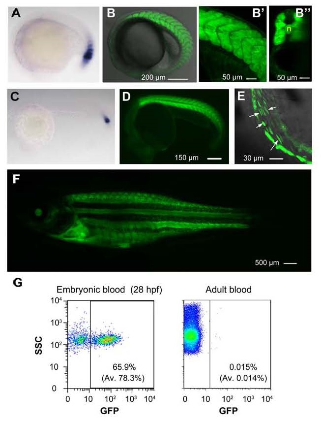
GFP expression pattern in Tg(ripply1:gfp) transgenic line. (A and C) Detection of ripply1 expression in nascent somites of wild-type embryos at 22s stage (A) and at 24 hpf (C) by in situ hybridization. (B-B′′) Confocal image of GFP expression in somites of a Tg(ripply1:gfp) embryo at 22s stage (B). The enlarged trunk region (B′) and an optical cross section (B′′) were shown. n, notochord. (D) GFP expression in transgenic embryos at 24 hpf. (E) GFP+ circulating hematopoietic cells (indicated by arrows) in the heart were observed by confocal microscopy at 36 hpf. See also Movie S1. (F) GFP was still expressed in adult fish. (G) Flow cytometry results showing the existence of GFP+ blood cells in the embryonic circulation at 28 hpf (left) and in the circulation of adults (right). Embryonic blood cells were sucked from the hearts of 5-10 embryos at 28 hpf. Adult blood cells were taken from the heart. The showed were representative results with average (Av) from 3 independent experiments in parenthesis.