
|
Fig. S1
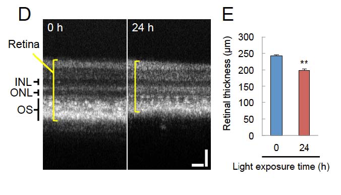
Optical coherence tomography (OCT) imaging of light damaged retina. (A) OCT image of a retina without light irradiation at 24 h. (B) Quantification of retinal thickness using OCT imaging results. For OCT imaging, fish were anesthetized for five minutes prior to imaging with 0.1% phenoxyethanol solution for about 1 min. Fish were laid with their right eye to the top and wrapped with a soft paper soaked with 0.1% phenoxyethanol (with the exception of the head). Before imaging, the pupils were dilated with 0.5% tropicamide (Santen, Osaka, Japan) and the eyes were lubricated with hydroxyethyl cellulose (Senju, Osaka, Japan). Retinal fundus images were taken during the same session as corresponding OCT scans using the MicronIV fundus camera (Phoenix Research Labs, Pleasanton, CA, USA). Data are shown as means ±SEM (n = 4). **p < 0.01 (Student’s t-test). Scale bars = 30 µm. INL, inner nuclear layer; ONL, outer nuclear layer; OS, outer segment.