Fig. 8
- ID
- ZDB-IMAGE-160823-18
- Genes
- Antibodies
- Source
- Figures for de Preux Charles et al., 2016
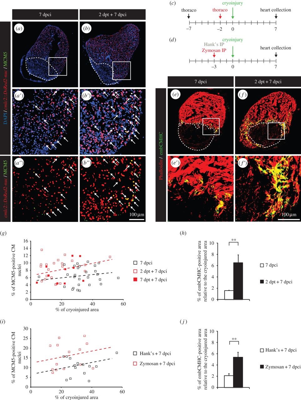
Fig. 8
Heart regeneration is improved in fish subjected to thoracotomy a few days before the cryoinjury. (a,b) Representative sections of hearts of transgenic cmlc2:DsRed2-nuc (red) fish labelled with the G1/S-phase marker MCM5 (green) at 7 dpci. Arrows indicate double-positive nuclei. (c,d) Experimental design. (c) Fish were subjected to thoracotomy at 7 or 2 days before the cryoinjury procedure. (d) Fish were injected intraperitoneally (IP) with Zymosan at 3 days before the cryoinjury procedure. (e,f) Representative sections of hearts labelled with the muscle marker Phalloidin (red) and embCMHC (green), a marker of undifferentiated CMs. (g,i) Scatter plot representing the percentage of MCM5-positive CM nuclei as a function of the cryoinjured area while using thoracotomy (g) (grey squares = 7 dpci; red squares = 2 dpt + 7 dpci; black squares = 7 dpt + 7 dpci) or Zymosan-IP (i) (grey squares = Hank′s + 7 dpci; black squares = Zymosan + 7 dpci) as preconditioning stimuli. (h,j) Quantification of the embCMHC-positive area after thoracotomy (h) or Zymosan-IP injection (j). A dashed line encircles the cryoinjured area (n ≥ 4 hearts; ≥2 sections per heart; **p < 0.01).

