
Fig. 7
- ID
- ZDB-IMAGE-160819-43
- Antibodies
- Publication
- Dupret et al., 2016 - The Polycomb Group Protein Pcgf1 Is Dispensable in Zebrafish but Involved in Early Growth and Aging
- All Figures
- Figures for Dupret et al., 2016
|
Fig. 7
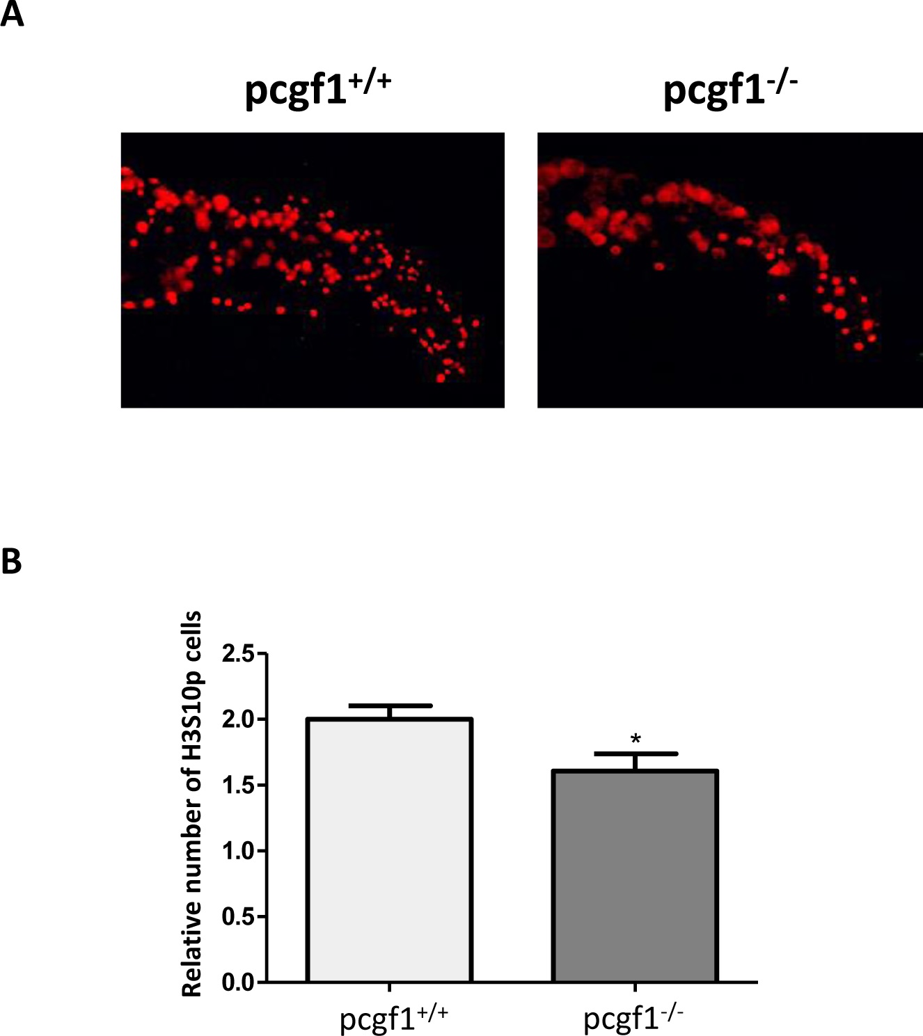
Cell proliferation measured by whole-mount immunohistochemistry using an anti-phosphohistone H3 antibody.
(A) Antibody staining against phosphorylated histone H3 (anti-H3S10p) in pcgf1+/+ and pcgf1-/- 24 hpf embryos. The caudal fin fold region of representative embryos is shown. (B) Quantification of H3S10p spots in the caudal fin fold expressed per unit of surface since pcgf1+/+ and pcgf1-/- embryos have different sizes at this developmental stage. Error bars indicate ± SD. Statistical significance was assessed by Student t-test analysis and significance expressed as the value of p and * indicates p < 0.01.