
Fig. 3
- ID
- ZDB-IMAGE-160816-3
- Genes
- Publication
- Nord et al., 2016 - Pax7 is required for establishment of the xanthophore lineage in zebrafish embryos
- All Figures
- Figures for Nord et al., 2016
|
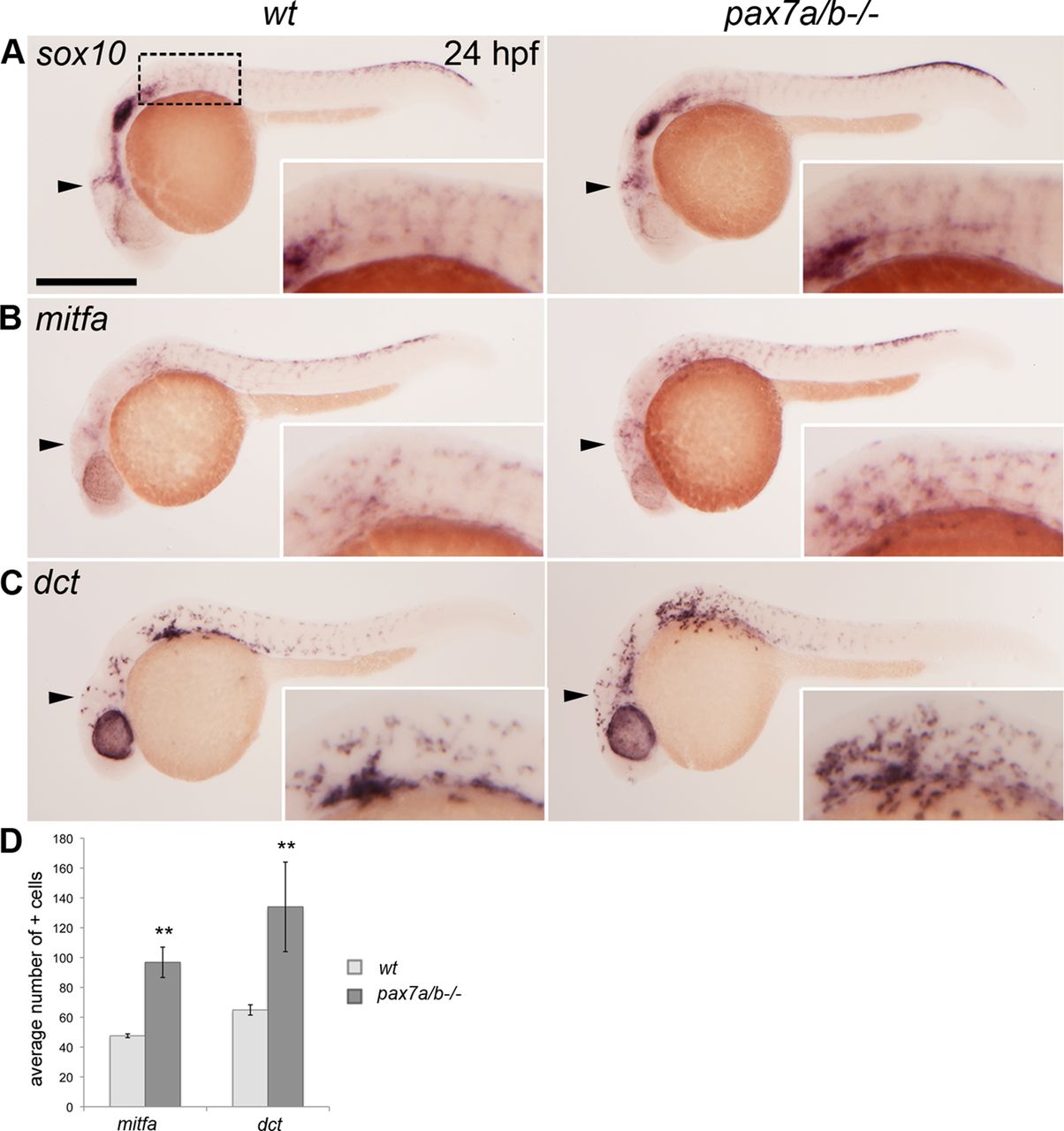
Fig. 3 The pax7a/pax7b double mutants have an increased number of melanoblasts. Whole-mount in situ hybridization on wt siblings and pax7a/pax7b double-mutant zebrafish embryo at 24 hpf showing the expression of (A) sox10 (n > 50 for siblings and n = 7 for pax7a/pax7b double mutants), (B) mitfa (n = 5 and 5), and (C) dct (n = 7 and 3). (D) Average number of mitfa+ and dct+ cells in the region anterior to the first somite on one side of wt siblings and pax7a/pax7b double-mutant embryos at 24 hpf; positive cells in the eye were excluded. Student’s t test was used to calculate significance; **p < 0.01. Error bars indicate SEM. Box indicates area of enlargement visualized in insets. Arrowheads indicate head region where changes in expression can be detected. Scale bar, 200 μm.