
IMAGE
Fig. 10
- ID
- ZDB-IMAGE-160728-4
- Genes
- Publication
- McCarthy et al., 2016 - An Fgf-Shh signaling hierarchy regulates early specification of the zebrafish skull
- All Figures
- Figures for McCarthy et al., 2016
Image
|
Figure Caption
Fig. 10
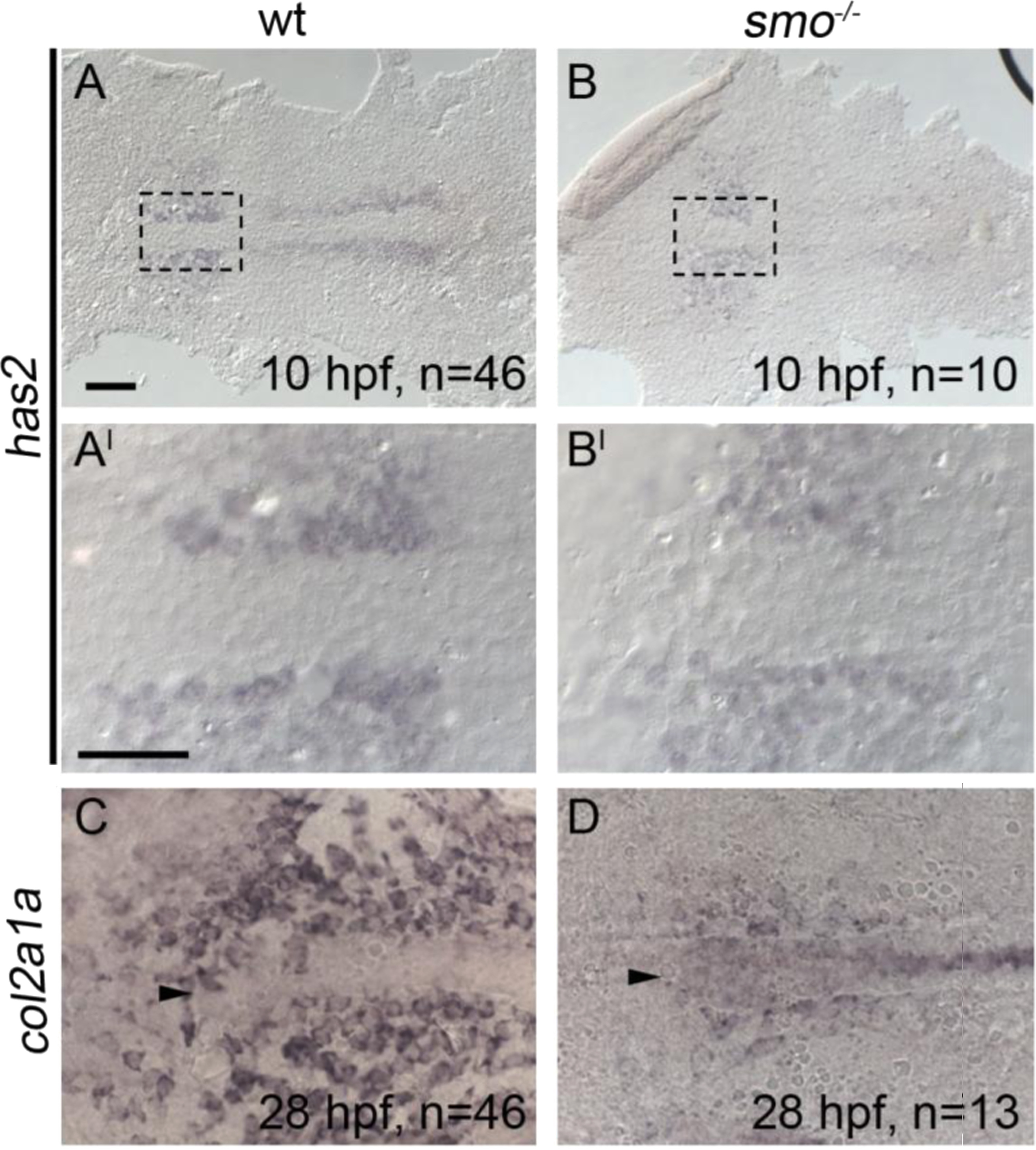
Chondrocyte differentiation is particularly sensitive to disruption of Hh signaling. All panels are anterior to the left. (A-B, A′-B′) At 10 h post-fertilization (hpf), the early mesoderm marker has2 is expressed in the anterior region of smoothened mutants and wildtypes (Compare B to A, B′ to A′). (C and D) However, smoothened mutants display a marked reduction in col2a1a expression in the forming postchordal neurocranium at 28 hpf compared to siblings (compare D to C, arrowhead denotes anterior notochord). Scale bar=50 µm in A and 10 µm in A′.
Figure Data
Acknowledgments
This image is the copyrighted work of the attributed author or publisher, and
ZFIN has permission only to display this image to its users.
Additional permissions should be obtained from the applicable author or publisher of the image.
Reprinted from Developmental Biology, 415(2), McCarthy, N., Sidik, A., Bertrand, J.Y., Eberhart, J.K., An Fgf-Shh signaling hierarchy regulates early specification of the zebrafish skull, 261-77, Copyright (2016) with permission from Elsevier. Full text @ Dev. Biol.