
|
Fig. S4
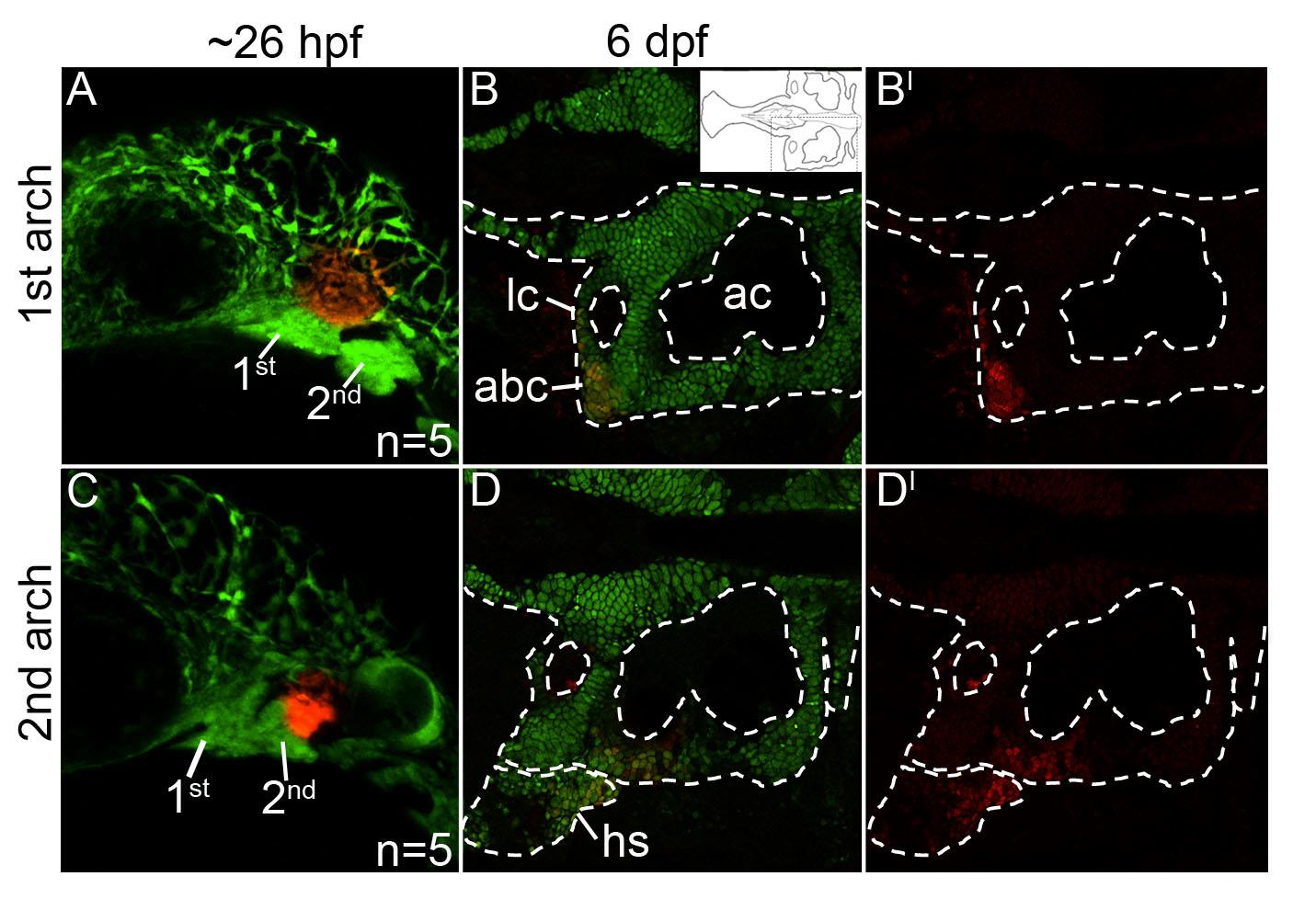
Mandibular- and hyoid-specific neural crest contributions to the postchordal neurocranium. (A-D′) Confocal images of sox10:kaede embryos at (A,C) 24 hours post fertilization and (B,B′, D, D′) 6 days post fertilization. Anterior is to the left in all images. (A) Photoconverted first arch neural crest cells, red fluorescence, contribute to the lateral anterior basicapsular commissure (B,B′, arrowhead, inset in B shows relative region shown in B and D). (C) Photoconverted second arch cells, red fluorescence, contribute to the lateral auditory capsule, with cells also labeled in the hyosymplectic, a second arch viscerocranial structure (D,D′). abc- anterior basicapsular commissure, ac- auditory capsule, hs- hyosymplectic, lc- lateral commissure.
Reprinted from Developmental Biology, 415(2), McCarthy, N., Sidik, A., Bertrand, J.Y., Eberhart, J.K., An Fgf-Shh signaling hierarchy regulates early specification of the zebrafish skull, 261-77, Copyright (2016) with permission from Elsevier. Full text @ Dev. Biol.