
Fig. 6
- ID
- ZDB-IMAGE-160727-50
- Genes
- Publication
- Hyatt et al., 1996 - Retinoic acid establishes ventral retinal characteristics
- All Figures
- Figures for Hyatt et al., 1996
|
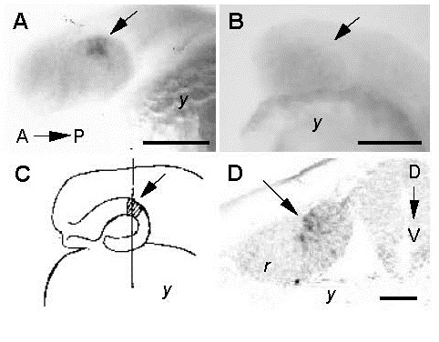
Fig. 6
In situ hybridization of msh[c] transcripts at 20 hpf in control embryos (A,D) and embryos treated with RA during the critical period (B). (A) In control embryos, msh[c] expression is localized to a small patch in the dorsal/posterior region of the eyecup (arrow) at 20 hpf. (B) No msh[c] expression is observed in the eyes (e) of treated embryos. (C) Illustration of a 20 hpf control embryo indicating the level of the transverse section (line D) shown in D. The arrow indicates the patch of msh[c] expression shown in A. (D) In transverse section, msh[c] expression (arrow) is found in the dorsal region of the neuroepithelium of the retina (R) in sections through the posterior region in the eyes of control embryos. D-V, dorsal-ventral axis; A-P, anterior-posterior axis; bar in A and B, 180 µm; bar in D, 50 µm.