
|
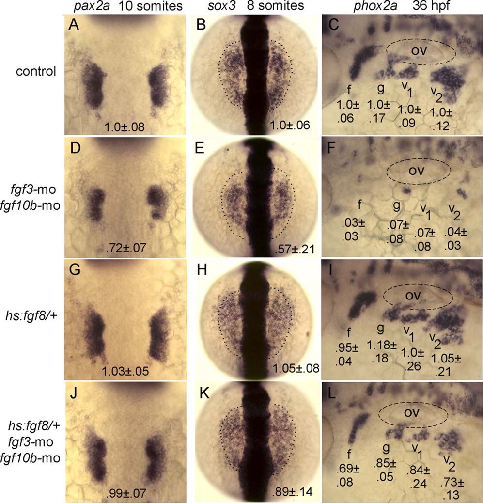
Fig. 8
Rescue of epibranchial development in fgf3-fgf10b deficient embryos by timed misexpression of fgf8. A-L: Dorsal views (anterior to the top) of pax2a expression at the 10-somite stage and sox3 expression at the 8-somite stage (normal boundaries of the control are outlined), and lateral views (anterior to the left) of phox2a expression at 36 hpf in embryos incubated at 35°C from 10 hpf until the eight-somite stage (for sox3) or the 10-somite stage (for pax2a and phox2a). Genetic manipulations are indicated along the left side. Positions of the otic vesicle (ov) and facial (f), glossopharyngeal (g), and vagal ganglia (v1 and v2) are indicated. Mean surface areas (± standard deviation, n ≥ 8), normalized to wild-type control embryos, are indicated for each structure