
Fig. 4
- ID
- ZDB-IMAGE-160726-4
- Genes
- Publication
- Heckel et al., 2015 - Oscillatory Flow Modulates Mechanosensitive klf2a Expression through trpv4 and trpp2 during Heart Valve Development
- All Figures
- Figures for Heckel et al., 2015
|
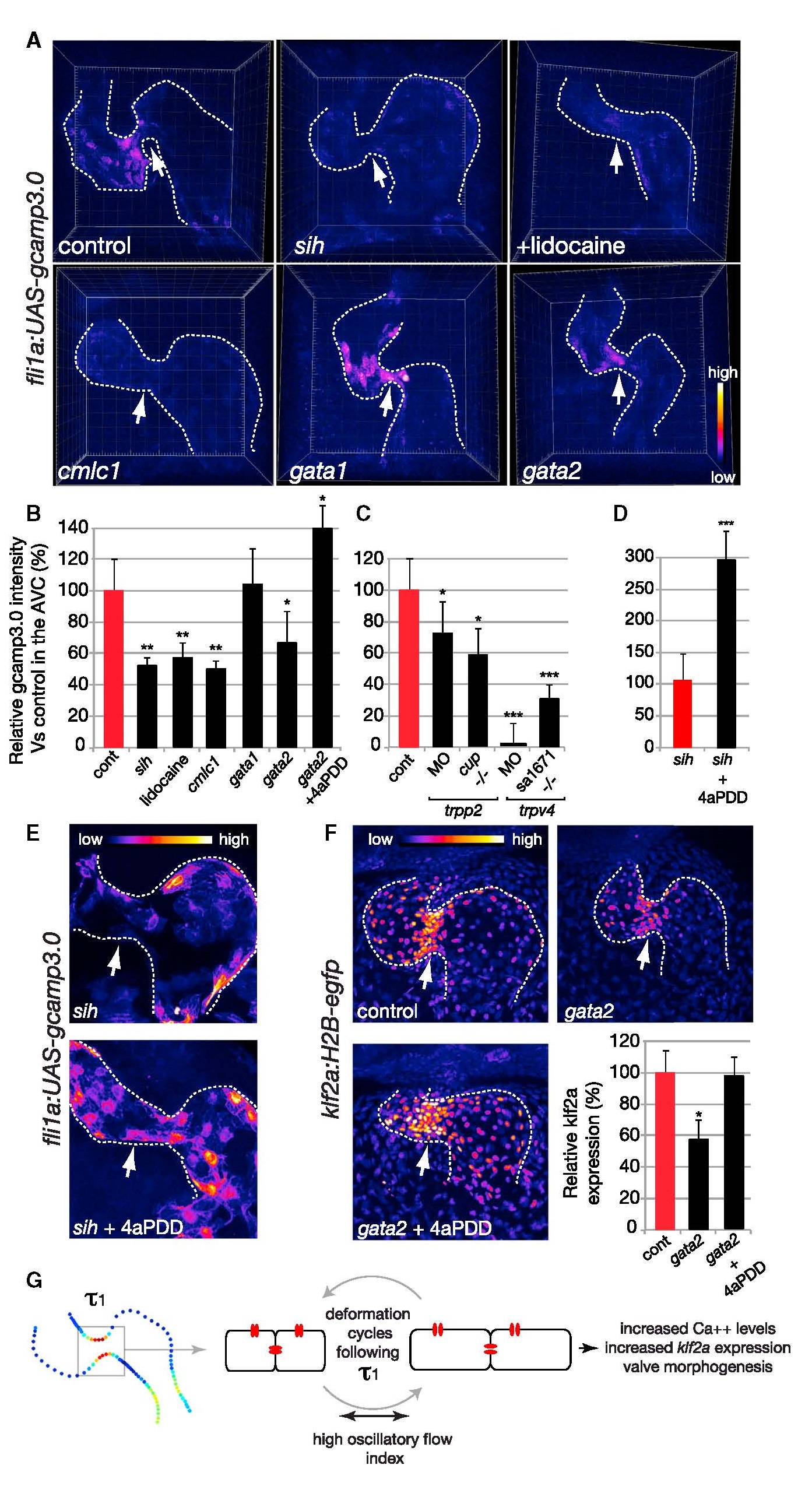
Fig. 4
Endocardial Intracellular Calcium Level Correlates with the Intracardiac FH Index and Depends on TRPs Channels Function
(A and B) Calcium level quantification in the embryonic heart of controls (n = 10), mutants of heart contraction (cmlc1 knockdown; n = 3), lidocaine-treated embryos (n = 3), gata1 (n = 3), gata2 (n = 6) MO, and gata2 MO plus 4α-PDD (n = 2). At 48 hpf, fishes with a reduced oscillatory flow index (sih, lidocaine, cmlc1, and gata2) show a significant reduction of intracellular calcium intensity in the AVC (white arrow), whereas the specific ligand of TRPV4 (4α-PDD) channels restores an almost normal intracellular calcium intensity in the AVC of the gata2 MO. *p < 0.05; **p < 0.01 ANOVA. Error bars indicate the SD.
(C) Calcium level quantification in the embryonic heart of control, trpp2 MO (n = 3), trpp2cup-/- (n = 6), trpv4 MO (n = 6), and trpv4sa1671-/- (n = 2) embryos. Error bars indicate the SD.
(D) 4α-PDD induces an increase in intracellular calcium in the absence of flow in the Tg(fli1a:gcamp3.0), sih (n = 5) hearts. ***p < 0.001 t test. Error bars indicate the SD.
(E) Confocal micrograph showing the effect of 4α-PDD on calcium signaling in sih.
(F) Confocal micrograph and intensity quantification of klf2a:H2B-eGFP intensity in gata2 knockdown with and without 4α-PDD treatment (n = 9 and n = 3, respectively). Error bars indicate the SD.
(G) Schematic highlighting a potential mechanism for oscillatory flow mechanodetection by the TRP channels (in red) specifically in the AVC.