
|
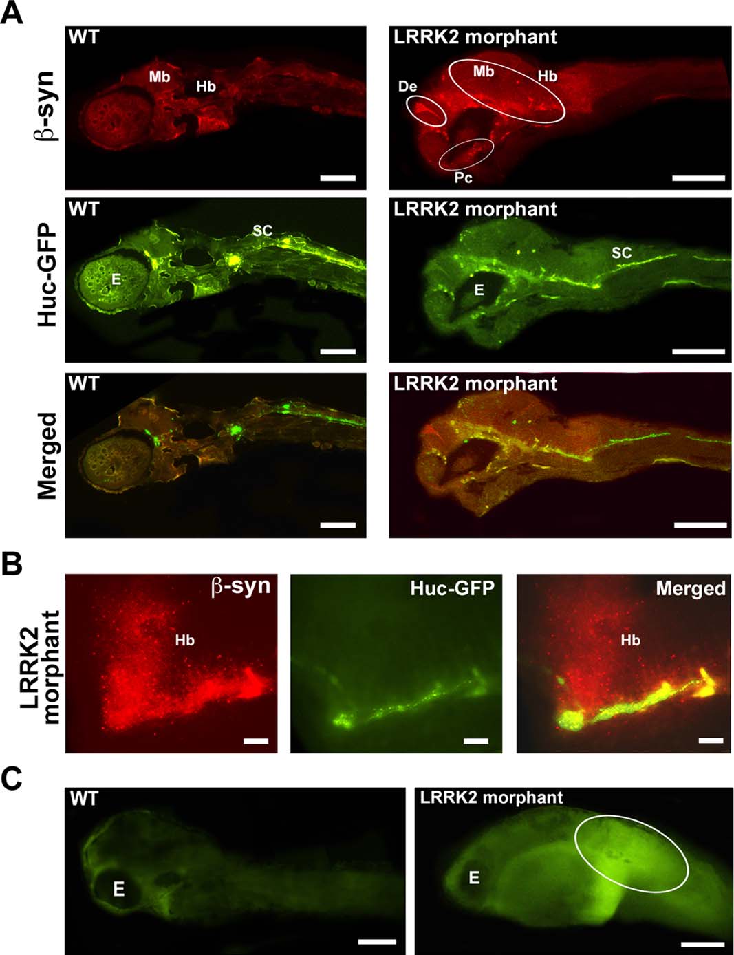
Fig. 8
Decreased LRRK2 level causes β-synuclein aggregation. A: Fluorescence microscopy images of 10-µm sections of 48 hpf wt embryos injected with HuC-GFP (green) and LRRK2 morphants coinjected with HuC-GFP (green) and the LRRK2 translational MO stained using the anti-β-synuclein antibody. β-Synuclein aggregates (red) were observed near the diencephalon (De), midbrain (Mb), hindbrain (Hb), and postoptic commissure (Pc) of the LRRK2 morphants but not in wt embryos. E, eye; De; diencephalon; Mb, midbrain; Hb, hindbrain; Pc, postoptic commisure. B: High levels of β-synuclein aggregates (red) were detected in the hindbrain region of the LRRK2 morphants. C: ROS staining of 72 hpf wt and LRRK2 morphant embryos showing high ROS levels in the tail region surrounding the bend in the LRRK2 morphant (oval) compared with wt embryos. Scale bars = 100 µm in A,C; 250 µm in B.