Fig. 4
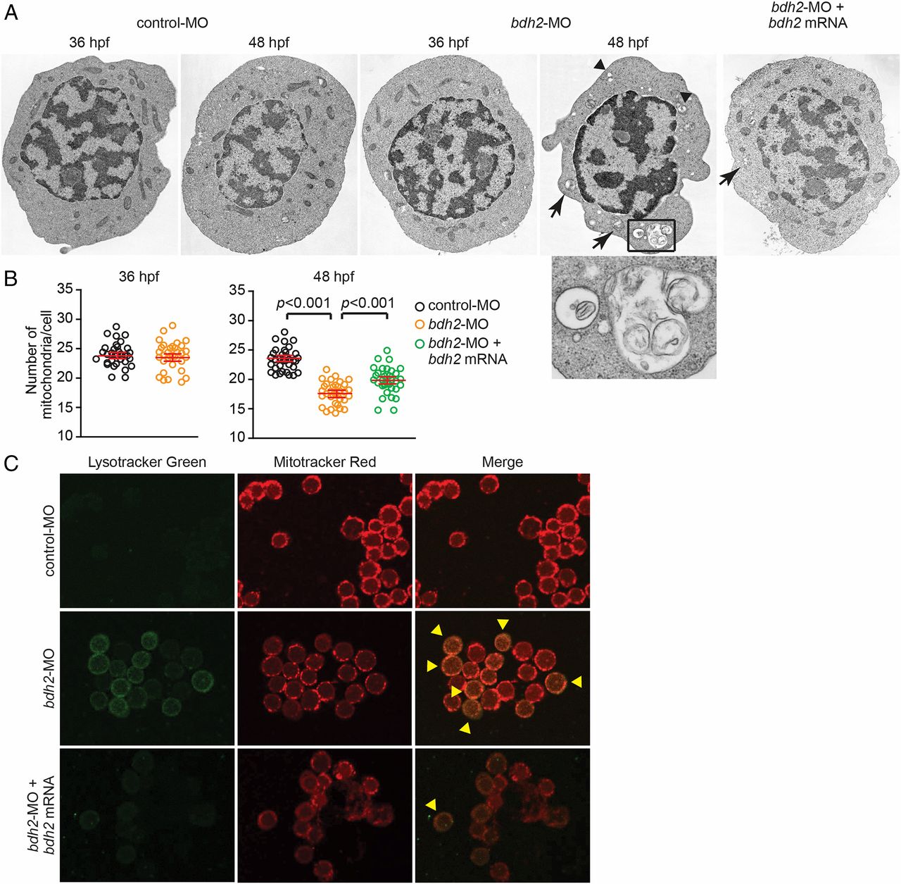
Bdh2-inactivated erythrocytes contain mitochondria in autophagosomal vesicles. (A) Representative EM images of erythrocytes isolated from control and bdh2 morphants. Arrows indicate autophagosomal vesicles. Arrowheads indicate lysosomal vesicles. (Scale bars: 1 µM.) Area in rectangle is enlarged. (Scale bar: Inset, 100 nM.) (B) Total mitochondria count per cell counted by EM. Data are mean ± SD for 50 cells. P < 0.05 was considered significant. (C) Confocal fluorescent imaging of LysoTracker- and MitoTracker-stained erythrocytes isolated from control and bdh2 morphants or bdh2-overexpressing bdh2 morphants. Arrowheads indicate costained cells. Representative images are depicted.

