
IMAGE
Fig. 3
- ID
- ZDB-IMAGE-160608-8
- Genes
- Publication
- Tokumasu et al., 2016 - ADAM12-deficient zebrafish exhibit retardation in body growth at the juvenile stage without developmental defects
- All Figures
- Figures for Tokumasu et al., 2016
Image
|
Figure Caption
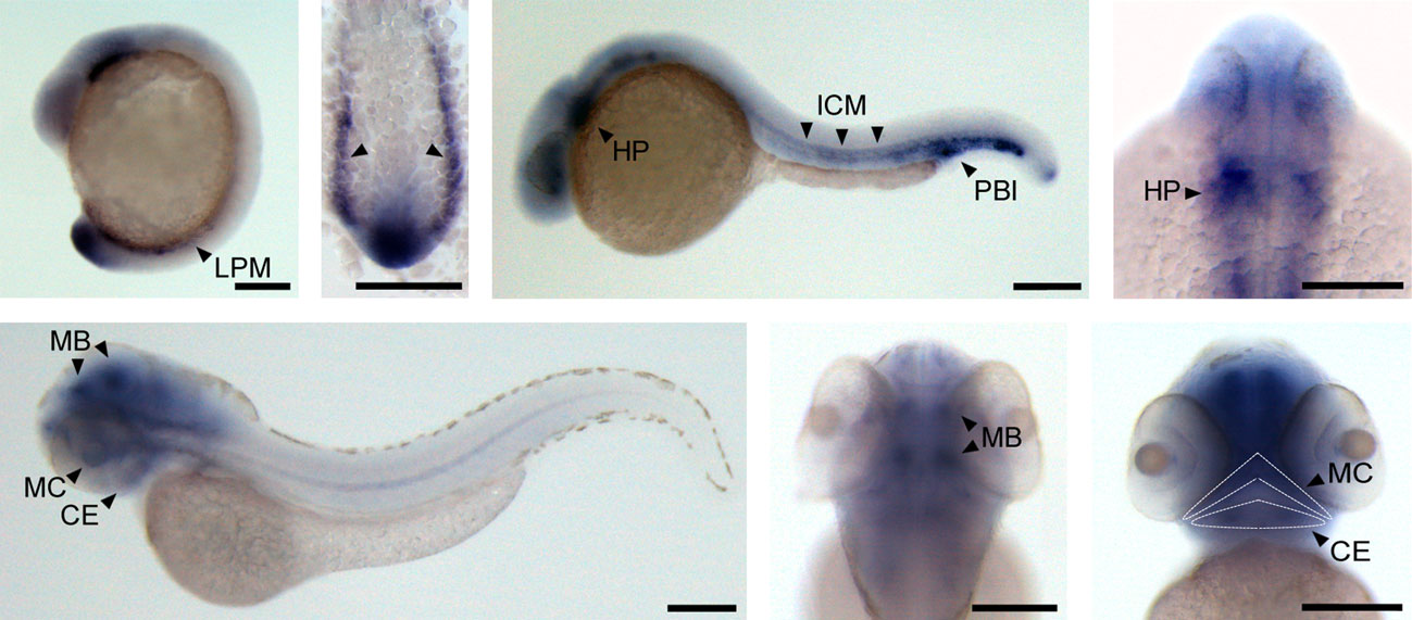
Fig. 3
Whole-mount in situ hybridization of zebrafish adam12. Micrographs of adam12 expression in zebrafish embryos, as determined by whole mount in situ hybridization at 16-hpf (A), 24-hpf (B), and 3-dpf (C). A′ shows a dorsal view of the posterior LPM; B′ shows a dorsal view of the heart primordium; C′ shows a dorsal view of the cephalic nerve; and C′′ shows a ventral view of the lower jaw. CE, ceratohyal; HP, heart primordium; ICM, intermediate cell mass; LPM, lateral plate mesoderm; MB, midbrain; MC, Meckel′s cartilage; PBI, posterior blood island. Scale bar: 200 µm.
Figure Data
Acknowledgments
This image is the copyrighted work of the attributed author or publisher, and
ZFIN has permission only to display this image to its users.
Additional permissions should be obtained from the applicable author or publisher of the image.
Full text @ Dev. Growth Diff.