
IMAGE
Fig. 5
- ID
- ZDB-IMAGE-160608-10
- Genes
- Antibodies
- Publication
- Tokumasu et al., 2016 - ADAM12-deficient zebrafish exhibit retardation in body growth at the juvenile stage without developmental defects
- All Figures
- Figures for Tokumasu et al., 2016
Image
|
Figure Caption
Fig. 5
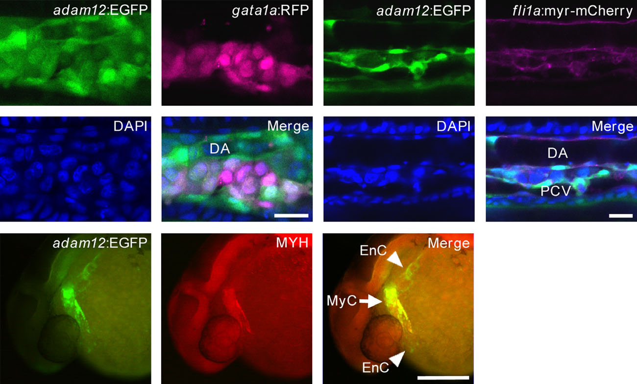
Identification of adam12-EGFP positive cells. (A, B) Confocal micrographs of the ICM region of double transgenic Tg[adam12:EGFP/gata1a:mRFP] or Tg[adam12:EGFP/fli1a:myr-mCherry] embryos. (A) Primitive erythroblasts expressing RFP at 24 hpf. (B) Endothelial cells expressing mCherry at 30-hpf. Nuclei are labeled with DAPI. DA, dorsal aorta; PCV, posterior cardinal vein. Scale bar: 20 µm. (C) MYH expression as determined by immunocytochemistry. MyC, myocardium; EnC, endocardium. Scale bar: 200 µm.
Figure Data
Acknowledgments
This image is the copyrighted work of the attributed author or publisher, and
ZFIN has permission only to display this image to its users.
Additional permissions should be obtained from the applicable author or publisher of the image.
Full text @ Dev. Growth Diff.