
Fig. 3
- ID
- ZDB-IMAGE-160524-60
- Publication
- Connors et al., 1999 - The role of tolloid/mini fin in dorsoventral pattern formation of the zebrafish embryo
- All Figures
- Figures for Connors et al., 1999
|
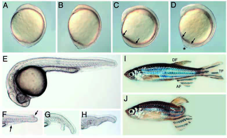
Fig. 3
Morphological defects visible in live mini fin mutant embryos. (A,B) 1-somite-stage mfn mutant (B) displays an oblong shape relative to wild type (A). 5- somite-stage wild-type (C) and mfn (D) mutant embryos. Arrows denote the tail bud length and the asterisk marks a protrusion of the tail bud seen in the mutant (D). 24 hpf wild type (E) and tails of mfn mutants exhibiting (F) a partial loss of tail fin (between the arrows) that extends to the dorsal side, (G) partial loss of the ventral fin with a bifurcation in the tail, (H) near complete absence of ventral fin and a kink in the tail. This occasionally observed kink is associated with a disruption in the notochord, which is also observed in the more strongly dorsalized mutant lost-a-fin (Solnica-Krezel et al., 1996, our unpublished observations). (I,J) mfn homozygous adult fish. Homozygous adult exhibiting a near wild-type appearance (I). Note that a pigmented stripe in the tail is disrupted in this mutant, a trait characteristic of mfn homozygous fish. Other adult mutants may display a partial or full (J) loss of their tail fin. All are lateral views. (A-D) dorsal to the right, (E-J) dorsal to the top. AF, anal fin; TF, tail fin; DF, dorsal fin.