
|
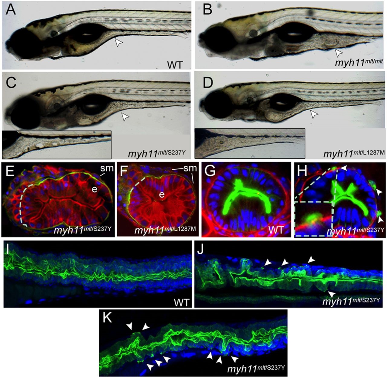
Fig. 2
Modifier mutations induce cell invasion in mlt heterozygotes. (A-D) Lateral views of live 6-dpf larvae. Arrowhead points to the mid-intestine. (A) A wild-type (WT) larva. (B) A homozygous mlt larva in which there is invasive expansion of the mid- and posterior intestine. (C) A mlt/S237Y compound heterozygote. Arrowhead points to a region of altered intestinal morphology. Inset shows a more severely affected mlt/S237Y compound heterozygote. (D) mlt/L1287M compound heterozygote. Mild and moderate (inset) phenotypes are shown as in C. (E,F) Histological section through the posterior intestine of 6-dpf mlt/S237Y and mlt/L1287M compound heterozygotes following their immunostaining with anti-Keratin (red) and anti-Laminin-1 antibodies (green). Dashed line indicates predicted position of basement membrane disrupted by the invasive epithelial cells; e, intestinal epithelium; sm, smooth muscle. (G,H) Histological section through the posterior intestine of 3-dpf Tg(miR194-Lifeact-GFP) wild-type and sibling mlt/S237Y larvae following immunostaining with anti-GFP (green) and anti-Laminin-1 (red) antibodies, showing GFP-labeled invadopodia (arrowheads) of epithelial cells extending through gaps in the basement membrane. Inset shows a higher-magnification view. (I-K) Confocal projections through the mid- and posterior intestine of 4-dpf Tg(miR194-Lifeact-GFP) wild-type and sibling mlt/S237Y (myh11mlt/S237Y) larvae following anti-GFP immunostaining (green). Arrowheads point to invadopodia protruding from the basal epithelial cell plasma membrane in the mlt/S237Y mutants. Blue, DAPI-stained nuclei.