
|
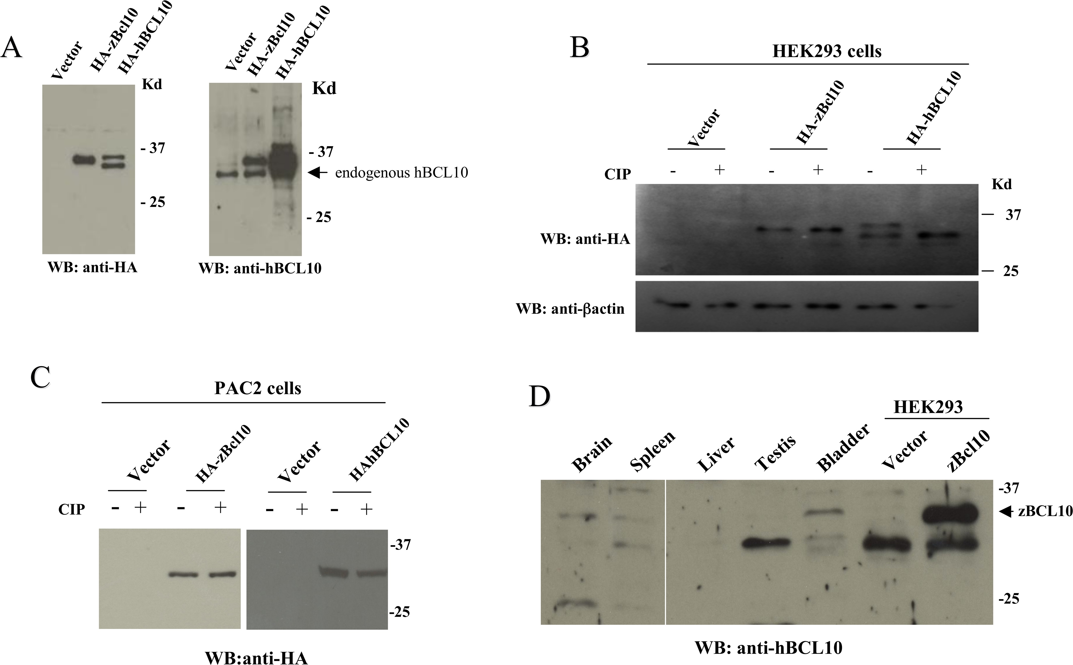
Fig. 2
A) Immunoblot analysis of lysates from HEK293 cells transfected with and expression vector empty (vector) or expressing HA-zBcl10 and HA-hBCL10. 24 hrs after transfection, cell lysates were prepared, separated by SDS/PAGE, and analyzed by immunoblot assay probed with anti-HA (left panel) and anti-BCL10 (right panel) antisera. An arrow indicates hBCL10 endogenously expressed by HEK293 cells. B) Cell lysates from HEK293 transfected cells were prepared as in A). Were indicated, before SDS/PAGE separation cell lysates were treated with 10 units of calf intestinal phosphatase (CIP) for 30 min at 37°C. C) The same experiment shown in B) was carried out in the embryonic zebrafish fibroblast cell line PAC2. D) Immunoblot analysis of proteic extracts from zebrafish organs probed with an anti-hBCL10 antisera. Cell extract from HEK293 cells transfected with zBcl10 was used as a positive control (arrow).