
|
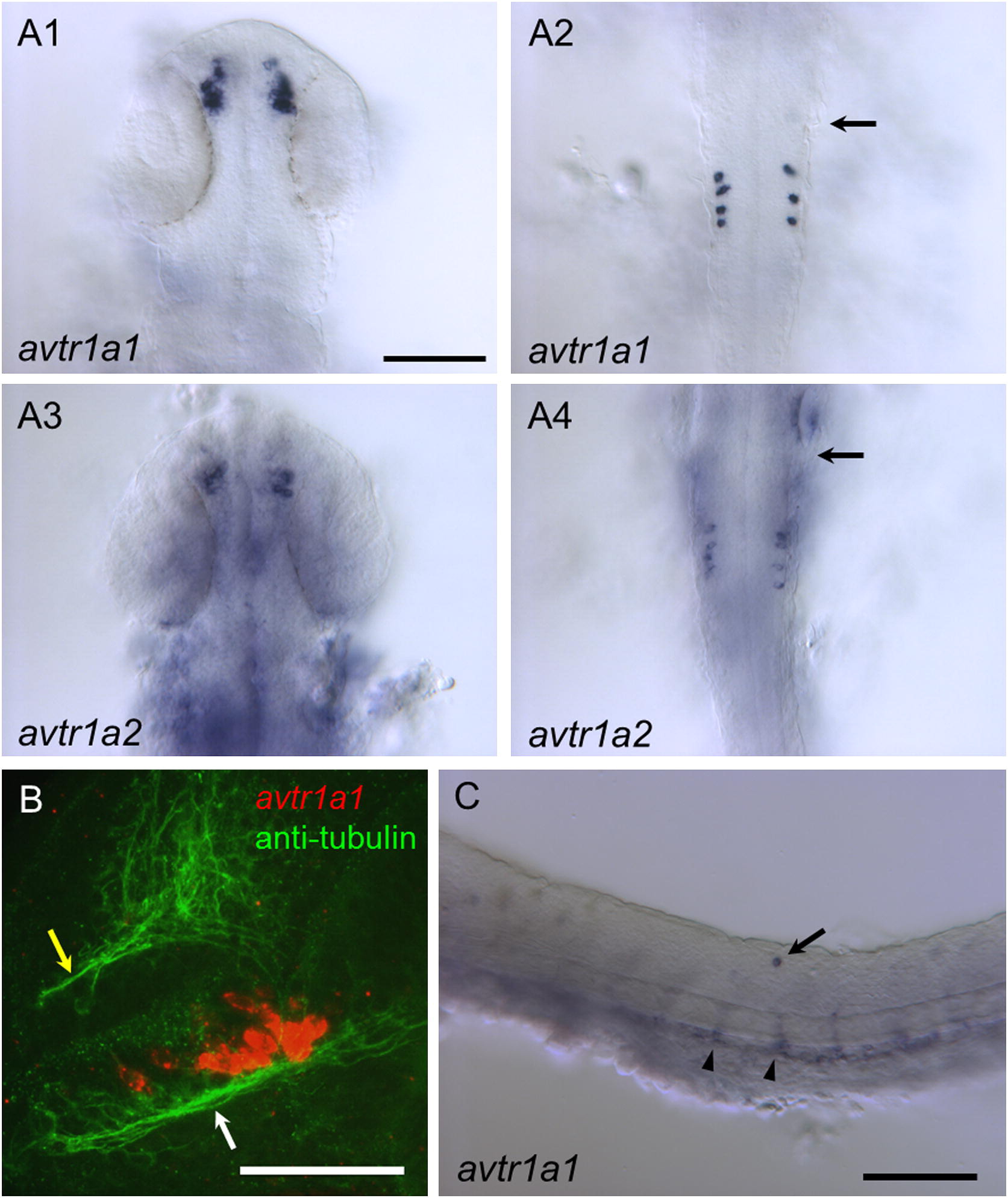
Fig. 2
Vasotocin receptors (avtr1a1 and avtr1a2) are expressed in discrete populations of cells in the CNS of 24/28 hpf embryos. (A) Dorsal views (anterior up) of in situ hybridizations of 25 hpf embryos showing that both avtr1a1 and avtr1a2 are expressed in the forebrain (A1, A3) and the posterior hindbrain (A2, A4). Arrows denote position of the posterior border of the otocyst. Scale:100 µm. (B) Combined in situ hybridization (red) and anti-acetylated α tubulin labeling of axons (green) showing avtr1a1 expressing neurons in the forebrain appear to project axons into the postoptic commissure (white arrow) and tract of the postoptic commissure (28 hpf, lateral view with anterior right and dorsal up). Yellow arrow denotes the anterior commissure. Scale: 50 µm. (C) Lateral view of trunk of 24 hpf embryo showing avtr1a1 expressing neuron in the dorsal spinal cord (arrow) and apparent endothelial cells forming blood vessels (arrowhead). Scale: 100 µm.
Reprinted from Gene expression patterns : GEP, 13(8), Iwasaki, K., Taguchi, M., Bonkowsky, J.L., and Kuwada, J.Y., Expression of Arginine Vasotocin Receptors in the Developing Zebrafish CNS, 335-42, Copyright (2013) with permission from Elsevier. Full text @ Gene Expr. Patterns