
IMAGE
Fig. 3
- ID
- ZDB-IMAGE-160420-4
- Genes
- Antibodies
- Publication
- Uribe et al., 2016 - A novel subset of enteric neurons revealed by ptf1a:GFP in the developing zebrafish enteric nervous system
- All Figures
- Figures for Uribe et al., 2016
Image
|
Figure Caption
Fig. 3
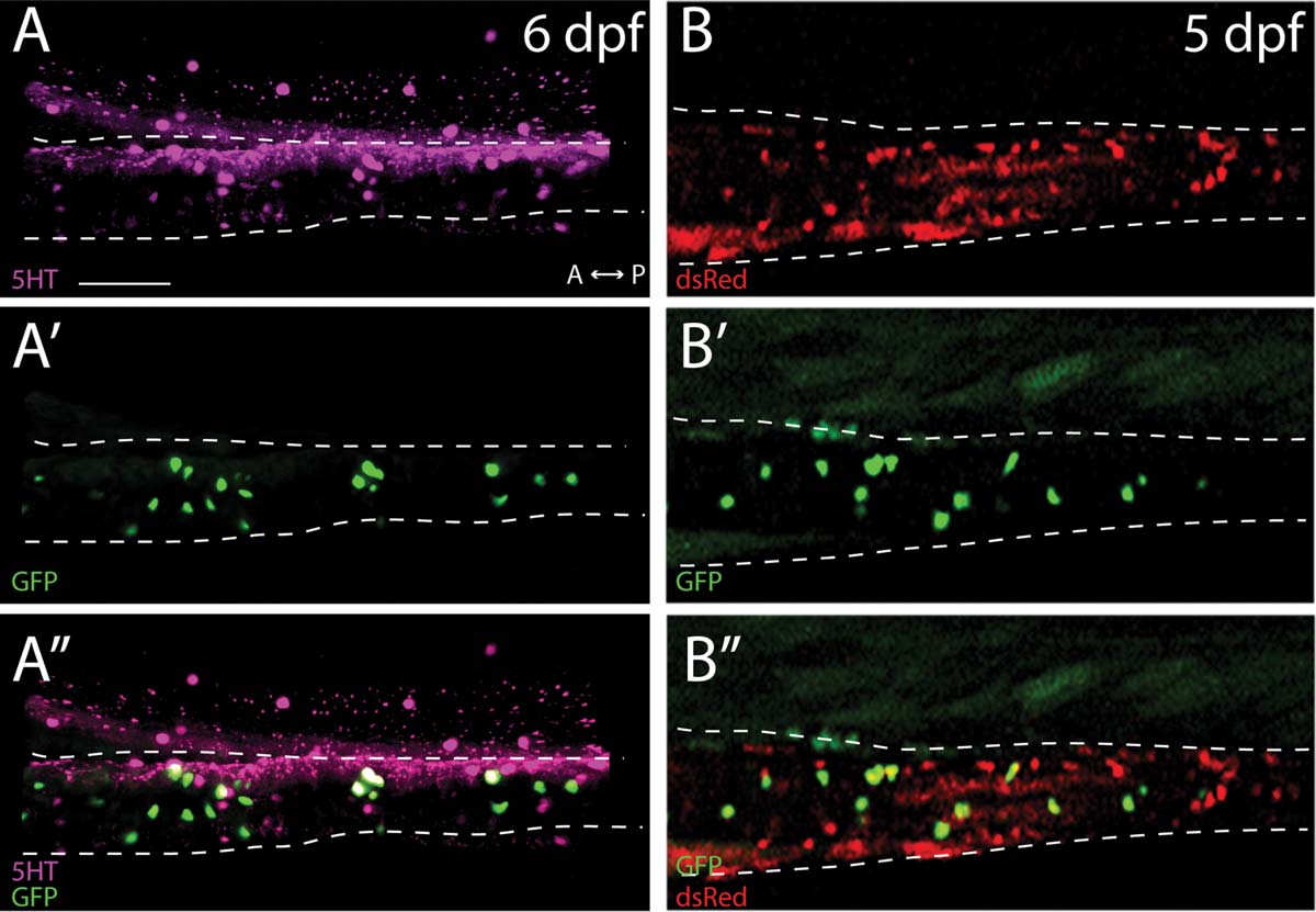
ptf1a:GFP cells are largely serotonergic enteric neurons that are derived from the neural crest. Maximum projection confocal stack of the midgut (A-A′′) reveal that ptf1a:GFP+ cells co-localize with the neurotransmitter 5HT at 6 dpf. (B-B′′) Images of live triple transgenic larval fish, ptf1a:GFP;-4725sox10:Cre;elf1a:loxp-GFP-loxp-dsRedpA, shows that ptf1a:GFP+ cells are neural crest derived. Scale bar: 70 µm.
Figure Data
Acknowledgments
This image is the copyrighted work of the attributed author or publisher, and
ZFIN has permission only to display this image to its users.
Additional permissions should be obtained from the applicable author or publisher of the image.
Full text @ Genesis