
IMAGE
Fig. S7
- ID
- ZDB-IMAGE-160407-19
- Publication
- Masuda et al., 2016 - ES1 is a mitochondrial enlarging factor contributing to form mega-mitochondria in zebrafish cones
- All Figures
- Figures for Masuda et al., 2016
Image
|
Figure Caption
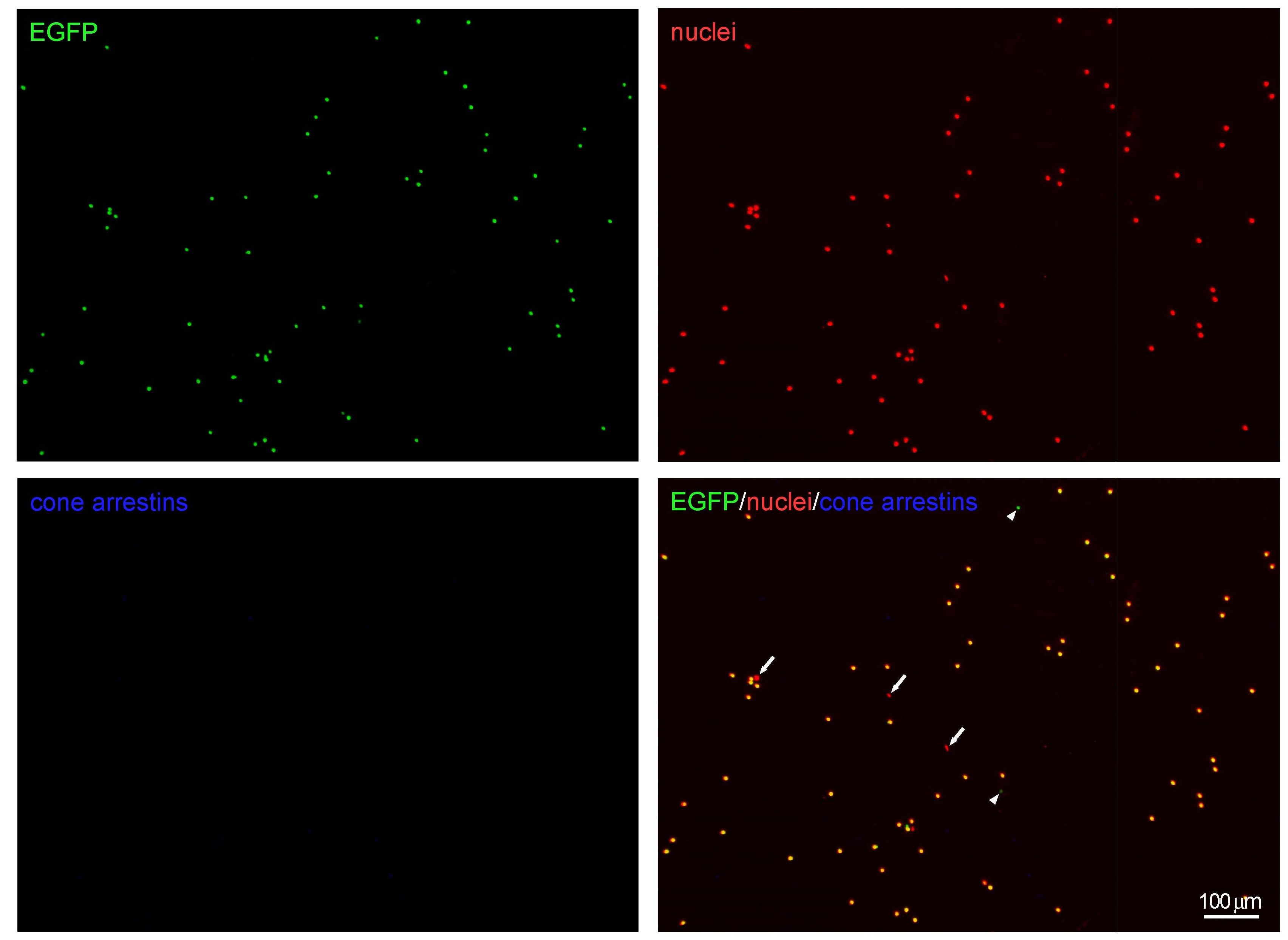
Fig. S7
Purity of FACS-purified rods. Representative microscopic images of a purified rod fraction from ES1-TG. Rod cell bodies were identified by signals for both EGFP (green) and nuclei (red, stained with DAPI). Approximately 97% of nuclei-containing dots were EGFP-positive. Few dots showed signal only for either EGFP (arrow heads) or nuclei (arrows), which were derived from rod outer segment or other cells, respectively. Possible contamination of cones was also investigated by immunostaining with antibodies against cone arrestins. Population of cone arrestins-immunopositive cells was only 0.28%.
Acknowledgments
This image is the copyrighted work of the attributed author or publisher, and
ZFIN has permission only to display this image to its users.
Additional permissions should be obtained from the applicable author or publisher of the image.
Full text @ Sci. Rep.