
IMAGE
Fig. S4
- ID
- ZDB-IMAGE-160407-16
- Publication
- Masuda et al., 2016 - ES1 is a mitochondrial enlarging factor contributing to form mega-mitochondria in zebrafish cones
- All Figures
- Figures for Masuda et al., 2016
Image
|
Figure Caption
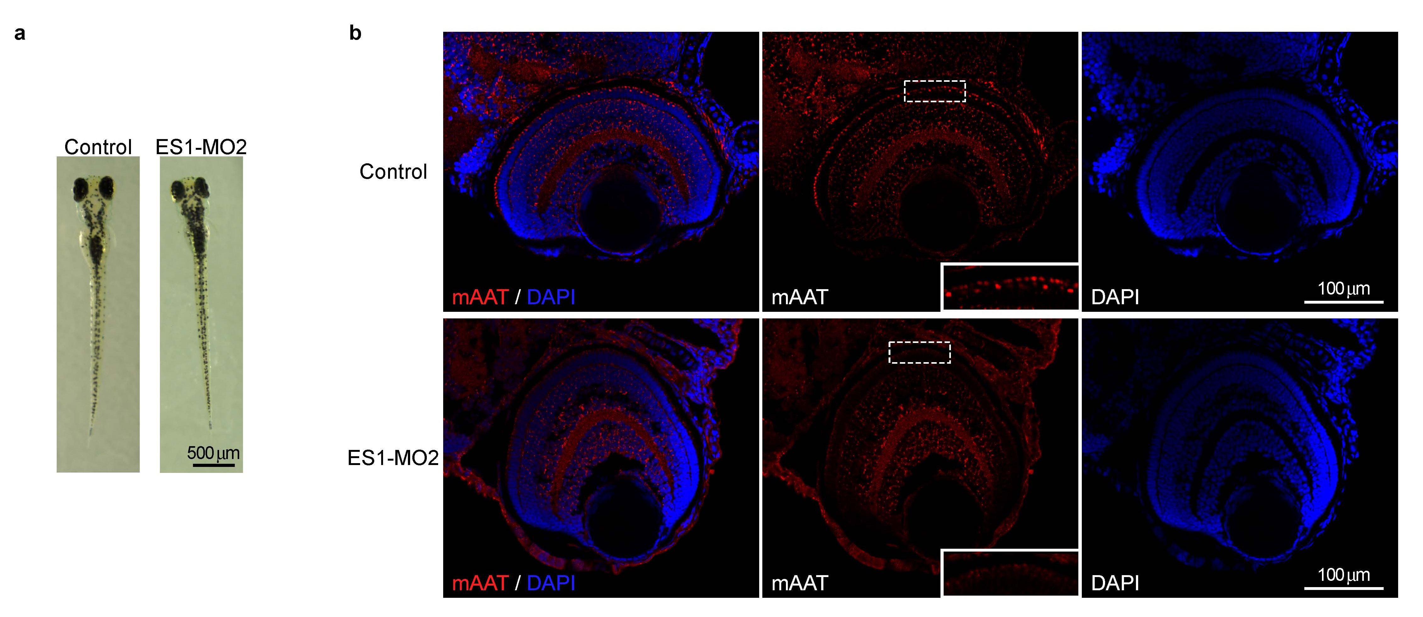
Fig. S4
Morphology of MO-injected larvae. (a) Representative whole body images of control MO- and ES1-MO2-injected 4 dpf larvae. (b) Immunohistochemistry of whole eye sections of control MO- and ES1-MO2-injected 4dpf larvae with mAAT antibody (red). Nuclei were stained with DAPI (blue). Immunopositive signals for mAAT were reduced only in cone ellipsoid layer (see dashed boxes and their magnified views shown in insets) of ES1- MO2-injected larvae compared to the control larvae.
Acknowledgments
This image is the copyrighted work of the attributed author or publisher, and
ZFIN has permission only to display this image to its users.
Additional permissions should be obtained from the applicable author or publisher of the image.
Full text @ Sci. Rep.