
|
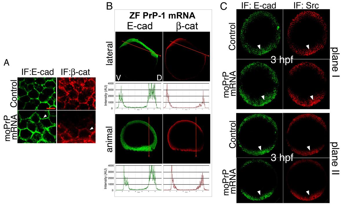
Fig. S5
Effects of embryonic PrP OE on the localization of SFKs and AJ components. A. Immunofluorescence in dorsal deep cells of 6 hpf embryos (animal views); arrowheads point at plasma membrane localization; scale bar = 10 µM. B. Gastrula midsections of 6 hpf ZF PrP-1 overexpressing embryos, immunostained as indicated and shown from lateral or animal perspectives. Graphs present fluorescence profiles along the ventral-dorsal axis (indicated by red arrows; V = ventral, D = dorsal). C. Blastula midsections of 3 hpf embryos (two different planes from animal view), immunostained as indicated; arrowheads point at areas of increased immunofluorescence.