
IMAGE
Fig. S1
- ID
- ZDB-IMAGE-160225-17
- Publication
- Fidelin et al., 2015 - State-Dependent Modulation of Locomotion by GABAergic Spinal Sensory Neurons
- All Figures
- Figures for Fidelin et al., 2015
Image
|
Figure Caption
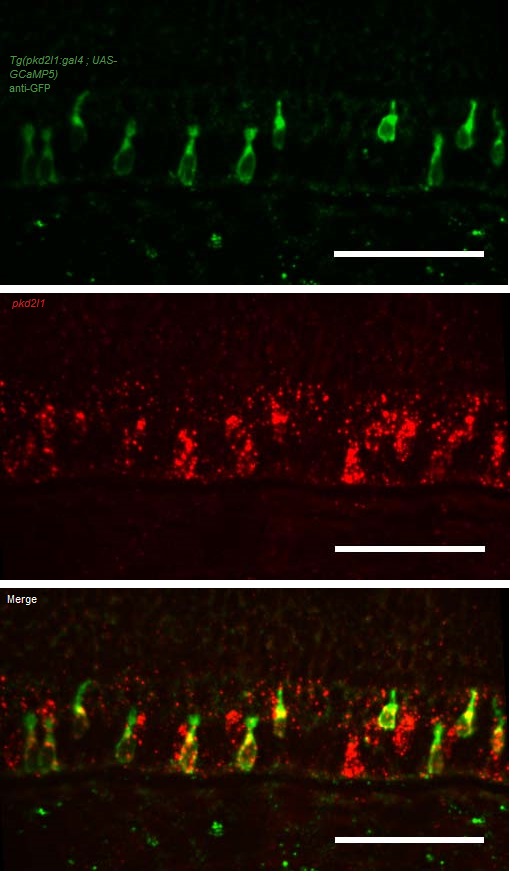
Fig. S1
Related to Figure 1. The Tg(pkd2l1:gal4)icm10 line drives expression in pkd2l1 positive cells in the zebrafish larva. Fluorescent in situ hybridization (FISH) for pkd2l1 was combined with an immunohistochemistry against GFP in a Tg(pkd2l1:gal4;UAS :GCaMP5G) larva at 3dpf. Spinal neurons expressing GCaMP5G (green) are positive for pkd2l1 (Pkd2l1+ in red), see arrowheads, n = 5 larvae. Scale bars represent 50 µm.
Acknowledgments
This image is the copyrighted work of the attributed author or publisher, and
ZFIN has permission only to display this image to its users.
Additional permissions should be obtained from the applicable author or publisher of the image.
Full text @ Curr. Biol.