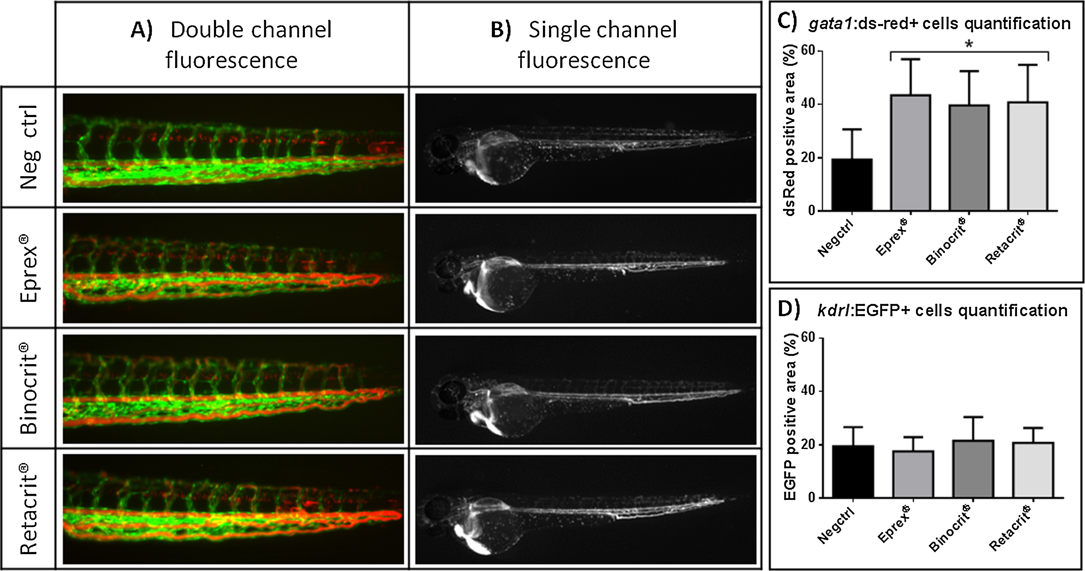
Fig. 4
Negative control (Neg ctrl), Eprex®, Binocrit® or Retacrit® injected in 48 hpf tg (kdrl:EGFP; gata1:ds-red) casper embryos and photographed after 4 h. (A) Double channel fluorescence: in green blood vessels, expressing kdrl:EGFP and in red circulating erythrocytes, expressing gata1:ds-red. Lateral views, anterior to the left, 11.5X magnification; (B) single red-channel fluorescence shows circulating erythrocytes, expressing gata1:ds-red. Lateral views, anterior to the left, 4X magnification. Quantification of (C) red fluorescence signal and (D) green fluorescence signal, measured with ImageJ 1.45 s image analysis software on a mean of 25 embryos for each experimental point. Asterisk indicates statistically significant increase of the positive area (p < 0.05), data are the mean ± S.D. of 3 independent experiments. (For interpretation of the references to color in this figure legend, the reader is referred to the web version of this article.)

